
- Title
-
Larval Zebrafish Lateral Line as a Model for Acoustic Trauma
- Authors
- Uribe, P.M., Villapando, B.K., Lawton, K.J., Fang, Z., Gritsenko, D., Bhandiwad, A., Sisneros, J.A., Xu, J., Coffin, A.B.
- Source
- Full text @ eNeuro
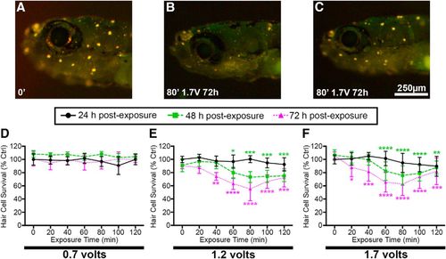
Acoustic stimulation results in exposure time-, intensity-, and post-exposure time-dependent reduction in DASPEI labeling, indicative of hair cell damage. A–C, Representative images of (A) unexposed larval zebrafish and (B, C) fish exposed to acoustic stimulation. Scale bar applies to all three images. Unexposed fish exhibit bright DASPEI staining indicative of a full complement of lateral line hair cells while fish exposed to 80 min of acoustic stimulation have diminished DASPEI labeling 72 h post-exposure. Two representative images of acoustically exposed fish are shown to depict the diversity of DASPEI labeling observed. D–F, Quantification of acoustic stimulation-induced hair cell loss. D, Fish exposed to 0.7 V show no reduction in DASPEI labeling. E, Fish exposed to 1.2 V of acoustic stimulation exhibit the greatest reduction in DASPEI labeling after 80 min of exposure and 72 h post-exposure. F, 1.7 V of acoustic stimulation produces similar DASPEI reduction to 1.2 V. Asterisks indicate significant differences from age-matched unexposed controls (*p < 0.05, **p < 0.01, ***p < 0.005, ****p < 0.001). Statistical analysis is shown in Table 1. N = 10–12 animals per treatment, values are mean ± SD. PHENOTYPE:
|
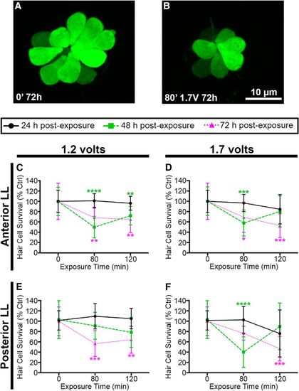
Acoustic stimulation decreases lateral line hair cell number. Unexposed (A) and acoustically stimulated (B) O2 neuromasts from myo6b:EGFP transgenic larval zebrafish. Scale bar applies to both images. 1.2 V (C) and 1.7 V (D) of acoustic stimulation significantly reduces the number of hair cells in five anterior lateral line neuromasts at 48 and 72 h after cessation of noise. 1.2 V (E) and 1.7 V (F) reduces hair cell number in pLL neuromasts P1 and P2. Asterisks indicate significant difference from age-matched unexposed controls (*p < 0.05, **p < 0.01, ***p < 0.005, ****p < 0.0001). Statistical analysis is shown in Table 1. N = 10–12 animals per treatment, values are mean ± SD. PHENOTYPE:
|
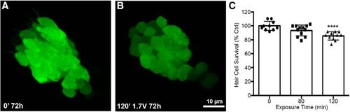
Acoustic stimulation produces an exposure time-dependent reduction in saccular hair cells. Unexposed (A) and acoustically stimulated (B) saccules from myo6b:EGFP transgenic zebrafish. There was no obvious spatial pattern to the damage in the acoustically exposed saccules. C, Treatment with 1.7 V of acoustic stimulation for 120 min significantly reduces saccular hair cell number when assessed 72 h post-exposure (one-way ANOVA; exposure time: F(2,30) = 11.89, p = 0.0002). Asterisks indicate significant difference from unexposed age-matched control (****p < 0.001). N = 10–12 animals per treatment, values represent mean ± SD. PHENOTYPE:
|
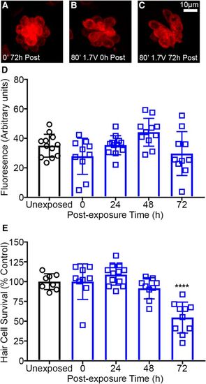
Loading of the mechanotransduction dependent dye FM 1-43FX is not affected by acoustic stimulation in wild-type *AB zebrafish. A–C, Representative images of neuromasts loaded with FM 1-43FX. Unexposed (A) and acoustically stimulated (B, C) neuromasts are brightly labeled with FM 1-43FX. D, Quantified FM 1-43FX fluorescence (normalized to hair cell number) is not significantly different in unexposed control versus 72 h post-exposure suggesting that acoustic stimulation does not alter hair cell mechanotransduction (one-way ANOVA; post-exposure time: F(4,50) = 4.001, p = 0.0068). Acoustically-exposed fish exhibit highly variable FM 1-43FX loading. E, Hair cell survival is reduced 72 h after acoustic stimulation (one-way ANOVA; post-exposure time: F(4,43) = 17.19, p < 0.0001). Hair cells were labeled with anti-parvalbumin and quantified in fixed animals. Asterisks indicate significant difference from unexposed control (****p < 0.001). N = 8–12 animals per treatment and values represent mean ± SD. PHENOTYPE:
|

ZFIN is incorporating published figure images and captions as part of an ongoing project. Figures from some publications have not yet been curated, or are not available for display because of copyright restrictions. |
|
ZFIN is incorporating published figure images and captions as part of an ongoing project. Figures from some publications have not yet been curated, or are not available for display because of copyright restrictions. PHENOTYPE:
|
|
ZFIN is incorporating published figure images and captions as part of an ongoing project. Figures from some publications have not yet been curated, or are not available for display because of copyright restrictions. PHENOTYPE:
|
|
ZFIN is incorporating published figure images and captions as part of an ongoing project. Figures from some publications have not yet been curated, or are not available for display because of copyright restrictions. PHENOTYPE:
|
|
ZFIN is incorporating published figure images and captions as part of an ongoing project. Figures from some publications have not yet been curated, or are not available for display because of copyright restrictions. PHENOTYPE:
|