
Fig. 1
- ID
- ZDB-FIG-181004-11
- Publication
- Severi et al., 2018 - Investigation of hindbrain activity during active locomotion reveals inhibitory neurons involved in sensorimotor processing
- Other Figures
- All Figure Page
- Back to All Figure Page
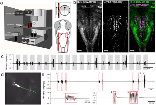
Single-cell resolution calcium imaging of the hindbrain during active visuomotor behavior. (a) The experimental set up consists of an in vivo 2-photon laser scanning microscope imaging at 980 nm and acquiring at 5.81 Hz while optomotor gratings are projected on a screen on the left or right side of the larva, which is embedded dorsal side up in agarose with the tail freed. A high-speed camera captures the tail movements from below at 250 Hz. The red box indicates the imaging region comprising the majority of the hindbrain. (b) Z-projection stack of several optical sections imaged from the dorsal view in Tg(HuC:GCaMP5G;glyt2:loxP-mCherry-loxP-Gal4; mifta−/−) transgenic larvae reveals the localization of GCaMP5G (left) in most neurons and mCherry (center) in glycinergic neurons (right, merge with GCaMP5G in green and mCherry in magenta). (c) Trials during which a single plane is imaged for 315 s with sequential presentations of moving optomotor stimuli for 10 s in open-loop (grey bars). Larvae initiate locomotion more frequently during the periods of stimulus motion than when the stimulus is stationary. (d) Single image extracted from a high-speed behavioral movie showing the larva under IR illumination with its head embedded in agarose and the tail cut free. Offline tracking of the tail in eight sections between the caudal end of the swim bladder and end of the tail. (e) Tail-bend angle (degrees) defined between the swim bladder and the end of the tail can be plotted and bouts of swimming are automatically extracted (red boxes). Tracking failures and obvious struggles were excluded from the dataset. Example swim bout expanded below (1) and example struggles expanded below (2,3). Red points mark automatically detected tail deflections. Each detected bout is bounded by a red box. Scale bars are 40 µm in (b). |