
Fig. 1
- ID
- ZDB-FIG-180921-26
- Publication
- Gistelinck et al., 2018 - Zebrafish type I collagen mutants faithfully recapitulate human type I collagenopathies
- Other Figures
- All Figure Page
- Back to All Figure Page
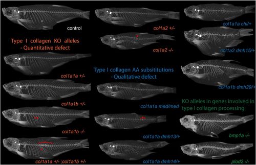
µCT images of different zebrafish models with affected type I collagen. For µCT scanning, we included several mutants with a nonsense or splice mutation in col1a1a, col1a1b, or col1a2, generating a knockout of these genes (col1a1a−/− not viable). These mutants are grouped together and indicated as “type I collagen knockout (KO) alleles—quantitative defect” (shown in orange). Another set of mutants carries mutations resulting in substitutions [indicated as “type I collagen amino acid (AA) substitutions—qualitative defect, shown in blue] of a Gly residue in α1(I) (col1a1admh13/+, col1a1admh14/+ and col1a1achi/+), α2(I) (col1a2dmh15/+), or α3(I) (col1a1bdmh29/+). The microwaved (col1a1amed/med) mutant carries a homozygous Glu substitution in α1(I). Finally, we included two mutant models with a knockout mutation in the bmp1a and plod2 genes (“KO alleles in genes involved in type I collagen processing,” shown in green). Representative fish from each mutant genotype are shown. Callus formation in ribs (arrowheads), local compressions of the vertebral column (brackets), and kyphosis (arrow), are indicated (also listed in Table 2). |
| Fish: | |
|---|---|
| Observed In: | |
| Stage: | Adult |