
Fig. 6
- ID
- ZDB-FIG-180921-30
- Publication
- Gistelinck et al., 2018 - Zebrafish type I collagen mutants faithfully recapitulate human type I collagenopathies
- Other Figures
- All Figure Page
- Back to All Figure Page
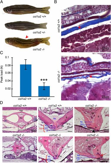
In-depth analysis of col1a2+/− and col1a2−/− mutant fish. (A) Visual phenotype of col1a2 mutant and wild-type siblings. Both col1a2+/+ control, and col1a2+/− mutant fish display a normal morphology, while col1a2−/− mutant are dysmorphic and display kyphosis (arrowhead). The typical stripe pattern in the skin is disturbed in col1a2−/− mutant fish, but normal in col1a2+/+ controls and col1a2+/− mutant fish. (B) Masson’s Trichrome staining of histological sections of the skin of an adult col1a2−/− mutant fish and a col1a2+/+ control sibling. Note the much thinner dermis (asterisk), composed of layers of collagen fibrils (stained blue) in col1a2−/− mutant fish compared with col1a2+/+ controls. (C) Average maximum tensile strength, measured by biomechanical testing of skin flaps dissected from five adult col1a2−/− mutant fish and five col1a2+/+ control fish, illustrating strongly decreased strength of soft connective tissues in mutant fish (P < 0.0001). (D) Histological analysis of the vertebral column of adult fish, sagittal sections stained with H&E. Shown at the Left are two adjoining vertebral body endplates (Vep, i.e., the region where two adjacent vertebral bodies meet and are connected by the IVL) in a col1a2+/+ control and a col1a2−/− mutant adult fish. The black rectangles indicate the region which is shown at a higher magnification in the image next to it on the Right, depicting the IVL in detail. In control fish, the IVL is typically composed of three distinct layers or tissues: the notochord sheet (Ns, mainly type II collagen), the outer elastin layer (OEL), and a layer of type I collagen fiber bundles (cI). In the col1a2−/− mutants, IVL composition is abnormal: the OEL was still present, the Ns layer could be observed but was extremely reduced, but no layer of type I collagen fibers (cI) could be observed. Col1a2−/− mutant bone (B) in the vertebral bodies near the Vep showed a loss of typical Sharpey fibers (Sf), while the notochord cells (Nc) displayed a loss of vacuoles, in addition to sclerosis (Sc). As shown in the two rightmost pictures, we observed fusions of adjacent vertebral bodies (black arrowhead) and dislocation of the IVL and Vep (red arrow) in some of the vertebrae, likely resulting in kyphosis seen in col1a2−/− mutant fish. Other abbreviations: Ep, epidermis; Mu, muscle fibers; Ob, osteoblasts; Scl, Scale. |
| Fish: | |
|---|---|
| Observed In: | |
| Stage: | Adult |