
Fig. S4
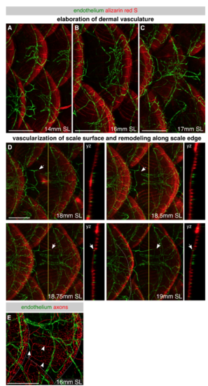
Development of skin vascularization, Related to Figure 4 (A-D) Lateral images of skin vasculature in fish expressing the pan-endothelial marker Tg(fli1a:EGFP) and stained with alizarin red S to label mineralized scales during late juvenile stages. Different individuals were imaged for each panel of A-C, whereas the same individual was repeatedly imaged in D. In panel A, note the large caliber vessels beneath each scale that connect to vessels beneath adjacent dorsal and ventral scales. In panels B and C, note the elaboration of the dermal network. In panel D, note that capillaries first extend along the radii above scales at ~18 mm SL. Arrows, vessel extending along scale surface. Yellow lines, planes of orthogonal sections. (E) Representative lateral view of the juvenile trunk in an animal expressing Tg(fli1a:EGFP) to label endothelium and Tg(p2rx3a>mCherry) to label axons. Arrow, tip of vessel growing along scale surface. Arrowheads, superficial scale nerves. Dashed lines, scale margins. Scale bars, 250 μm. |
Reprinted from Developmental Cell, 46(3), Rasmussen, J.P., Vo, N.T., Sagasti, A., Fish Scales Dictate the Pattern of Adult Skin Innervation and Vascularization, 344-359.e4, Copyright (2018) with permission from Elsevier. Full text @ Dev. Cell