
Fig. S3
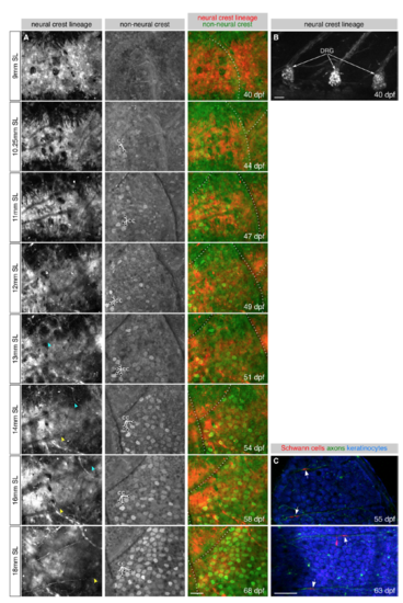
DRG axons innervate the epidermis during late juvenile stages, Related to Figure 2 (A) Repeated imaging of a single lateral skin region during scale formation. Arrowheads, individual axons extending along the scale surface. Dashed lines, scale margins. cc, club cells. (B) Lateral view of three dorsal root ganglia from the same fish as shown in panel A. (C) Isolated scales imaged at the indicated stages. Arrows, presumptive Schwann cells. Transgenes: (A) Tg(-28.5Sox10:Cre); Tg(ubb:GswitchR); (B) Tg(-28.5Sox10:Cre);Tg(ubb:GswitchR) (red channel only); (C) axons [Tg(p2rx3b:EGFP)], and keratinocytes and Schwann cells [Tg(-28.5Sox10:Cre);Tg(actb2:BswitchR)]. Scale bars, 50 μm. |
Reprinted from Developmental Cell, 46(3), Rasmussen, J.P., Vo, N.T., Sagasti, A., Fish Scales Dictate the Pattern of Adult Skin Innervation and Vascularization, 344-359.e4, Copyright (2018) with permission from Elsevier. Full text @ Dev. Cell