
Fig. 6
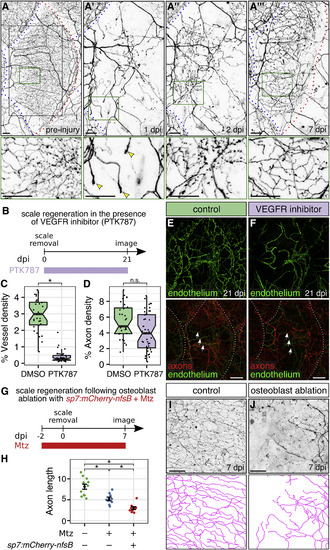
Osteoblasts, but Not Blood Vessels, Promote Epidermal Re-innervation following Injury (A) Regeneration of a single scale and associated axons. Black box, region of images in (A′)–(A‴). Green boxes, regions of magnification in lower panels. Red dashed lines, scale margins before and after removal. Blue dashed lines, margins of non-injured scales. Note that regenerating axons developed growth cones in the dermis at 1 dpi (arrowheads), sparsely innervated the epidermis at 2 dpi, and more extensively innervated the epidermis at 7 dpi. (B) Experimental design for scale regeneration in the presence of 500 nM PTK787. (C and D) Boxplots of vessel (C) and axon (D) density based on Tg(fli1a:EGFP) expression or acTubulin staining, respectively, in regenerating scales at 21 dpi. n = 31–44 scales from n = 2–3 fish. ∗p < 0.01; n.s., not significant (p > 0.01); Wilcoxon rank-sum test. (E and F) Representative lateral views of the indicated markers in control (DMSO) or VEGFR inhibitor (PTK787)-treated skin. Note that neither treatment prevented the formation or maintenance of nerves (arrows) over the course of the experiment. Dashed lines, scale margins. (G) Timeline of osteoblast ablation using metronidazole (Mtz). (H–J) Quantification (H) and representative lateral views and tracings (I and J) of regenerating axons in control or osteoblast-ablated fish (Mtz) at 7 dpi. Axon length is expressed as millimeters per 0.0625 mm2 region of skin. n = 9–12 regions/treatment from n = 2–3 fish. Black bars, mean ± SEM. ∗p < 0.01, Wilcoxon rank-sum test. Transgenes: (A) axons (Tg(p2rx3a>mCherry)); (E and F) axons (Tg(p2rx3a>mCherry)) and endothelium (Tg(fli1a:EGFP)); (I and J) axons (Tg(p2rx3b:EGFP)). Scale bars, 50 μm (A, I, and J) and 200 μm (E and F). See also Figure S5. |
| Gene: | |
|---|---|
| Fish: | |
| Condition: | |
| Anatomical Terms: | |
| Stage: | Adult |
| Fish: | |
|---|---|
| Conditions: | |
| Observed In: | |
| Stage: | Adult |
Reprinted from Developmental Cell, 46(3), Rasmussen, J.P., Vo, N.T., Sagasti, A., Fish Scales Dictate the Pattern of Adult Skin Innervation and Vascularization, 344-359.e4, Copyright (2018) with permission from Elsevier. Full text @ Dev. Cell