
Fig. 2
- ID
- ZDB-FIG-180913-53
- Publication
- Asakawa et al., 2018 - Protocadherin-Mediated Cell Repulsion Controls the Central Topography and Efferent Projections of the Abducens Nucleus
- Other Figures
- All Figure Page
- Back to All Figure Page
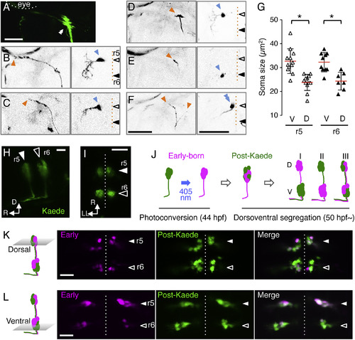
Soma Size Topography of mnr2b-ABNs (A) Left abducens nerve terminal of Tg[mnr2b-hs:Gal4]; Tg[UAS:GFP] larva at 5 dpf. (B–E) Axon terminals (orange) and somata (blue) of single abducens motor neurons. (F) Two neighboring abducens motor neurons on the dorsal side of r5, possessing either an en grappe-type (left arrowhead) or en plaque-type (right arrowhead) terminal, respectively. (G) The soma size of the dorsal and ventral cells in r5 and r6. N = 40 cells from 22 animals. ∗p < 0.05 (t test). (H and I) The lateral (H) and dorsal (I) views of an mnr2b-ABNs in the Tg[mnr2b-hs:Gal4]; Tg[UAS:Kaede] embryo at 44 hpf. (J) Birth-order labeling of mnr2b-ABNs and possible segregation patterns of early-born neurons along the dorsoventral axis (magenta in I, II, and III). Green fluorescence of Kaede that appears after photoconversion is indicated as post-Kaede. (K and L) Dorsal (K) and ventral (L) sections of r5 and r6 in the photoconverted Tg[mnr2b-hs:Gal4]; Tg[UAS:Kaede] larvae at 72 hpf. Scale bars indicate 50 μm in (A)–(F) and 20 μm in (H), (I), (K), and (L). |
| Genes: | |
|---|---|
| Fish: | |
| Anatomical Terms: | |
| Stage Range: | High-pec to Day 5 |