
Fig. 5
- ID
- ZDB-FIG-180912-97
- Publication
- Lozano-Ortega et al., 2018 - Hair cell identity establishes labeled lines of directional mechanosensation
- Other Figures
- All Figure Page
- Back to All Figure Page
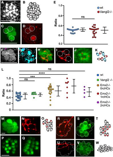
Regenerative synaptogenesis of neuromasts. (A–B) Apical aspect of a neuromast in Vangl2 mutant, labeled with fluorescent phalloidin (panel A) and its schematic representation (panel B). These images highlight the loss of coherent epithelial planar polarity in the neuromast. (C–D) Immunohistochemical staining of a horizontal neuromast in the transgenic line Et(krt4:EGFP)sqet4 mutant for Vangl2 showing HCs (panel C) and Emx2 (panel D). Dotted circles indicate Emx2(+) cells. (E) Quantification of Emx2 expression in wild-type (blue) and Vangl2 mutant (red) neuromasts. Numbers on the y-axis indicate the fraction (ratio) of the hair cells expressing Emx2; one-way ANOVA. Error bars indicate mean ± SD. (F–K) A Vangl2−/− neuromast expressing EGFP in hair cells (panel F), immunostained for Emx2 (panel G), labeled with 488-phalloidin to reveal planar polarity (panel J) and expressing mCherry in a single LAN (panel H–I). Emx2 is expressed in 3 out of 7 hair cells (dashed circles) (panel G). A purple star marks an Emx2(+) cell that is not a hair cell (panel F–G). The marked LAN (panel H–I) innervates the 4 Emx2(−) hair cells as shown in the scheme (panel K), which is color-coded (blue for Emx2[+] and grey for Emx2[−]). (L) Quantification of the number of hair cells innervated by an identified axon in horizontal neuromasts in wild-type specimens (blue) and in those lacking Vangl2 (green) or Emx2 (orange/red/violet). Emx2 mutant neuromast were separated into those containing 0 rcHCs (orange), 1 rcHC (red), and 2 rcHCs (violet). Numbers on the y-axis indicate the fraction (ratio) of innervated hair cells. ***p < 0.001; one-way ANOVA. Error bars indicate mean ± SD. (M–N) A neuromast in the transgenic line Et(krt4:EGFP)sqet4 expressing mCherry in a single LAN, showing HCs (panel M) and axonal terminal arborization (panel N). (O) A scheme of panel M–N indicating hair cell orientation (eccentric black dots) and the synapse of the individualized axon (red asterisks). (P–Q) A vertical (panel P) and horizontal (panel Q) neuromast from specimens lacking Emx2, showing the typical homogeneous polarization of hair cells. (R–S) A vertical neuromast lacking Emx2 in the transgenic line Tg[pou4f3:GAP-GFP; pou4f3:ctbp2l-mKOFP] expressing mCherry in a single LAN, showing juxtaposition of LAN neurites (red) with the active-zone Ribeye(+) puncta (yellow) (panel R), and presence of Ribeye(+) puncta (yellow) in hair cells (green) (panel S). Dotted line indicates the hair cells. (T) A scheme of panel R–S indicating hair cell orientation (eccentric black dots) and the synapse of the individualized axon (red asterisks). Note that the noninnervated hair cells are Ribeye(−) and therefore are likely immature. (U–V) A horizontal neuromast lacking Emx2 in the transgenic line Tg[pou4f3:GAP-GFP] expressing mCherry in a single LAN, showing no synapse of the LAN with any of the hair cells. Dotted line indicates the hair cells. (W) A scheme of panel U–V showing hair cell orientation (eccentric black dots). Scale bars are 10 μm. EGFP, enhanced green fluorescent protein; Emx2,; HC, hair cell; LAN, lateralis afferent neuron; ns, not significant; rcHC, rostrocaudal HC; Vangl2,; wt, wild-type. |
| Fish: | |
|---|---|
| Knockdown Reagent: | |
| Observed In: | |
| Stage Range: | Protruding-mouth to Days 7-13 |