
Fig. 4
- ID
- ZDB-FIG-180912-96
- Publication
- Lozano-Ortega et al., 2018 - Hair cell identity establishes labeled lines of directional mechanosensation
- Other Figures
- All Figure Page
- Back to All Figure Page
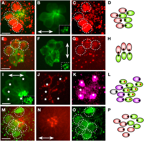
Emx2 expression in larval neuromasts. (A–C) Immunohistochemical staining of a horizontal neuromast in the transgenic line Tg[myo6b:actb1-EGFP] showing HCs (green) and Emx2 (red, dotted circles). Inset in panel B is a superficial image of the hair cells’ apices revealing their planar polarization. (D) Scheme of the neuromast in panel A–C indicating Emx2-expressing (red) and nonexpressing (green) hair cell. (E–G) Immunohistochemical staining of a vertical neuromast in the transgenic line Tg[myo6b:actb1-EGFP] showing HCs (green) and Emx2 expression (red, dotted circles). Inset in panel F is a superficial image of the hair cells’ planar polarization. (H) Scheme of the neuromast in panel E–G indicating Emx2-expressing (red) and nonexpressing (green) hair cell. (I–K) Immunohistochemical staining of a horizontal neuromast in the transgenic line Tg[myo6b:actb1-EGFP] showing HCs (green) (panel I), a single LAN expressing mCherry (red) (panel J). White asterisks indicate Emx2-expressing hair cells (purple) (panel K). Inset in panel I reveals hair cell planar polarization. (L) Scheme of the neuromast in panel I–K indicating Emx2-expressing hair cell (purple), nonexpressing hair cell (green), and the synapse of the individualized axon (red asterisks). The polarity of hair cell could not be determined (grey). In panel B, F, I, and N, neuromast orientation is indicated by a double-head arrow. (M–O) Staining of a horizontal neuromast in a neurogenin1 mutant specimen, with an antibody to Emx2 and phalloidin to reveal hair cell apices. It shows Emx2-expressing cells (green, dotted circles) and hair cell orientation (panel N) (red, double-head arrow) (panel N). (P) Scheme of the neuromast in panel M–O indicating Emx2-expressing (red) and nonexpressing (green) hair cell, indicating hair cell orientation (eccentric black dots). Scale bars are 10 μm. HC, hair cell; LAN, lateralis afferent neuron. |