
Fig. 3
- ID
- ZDB-FIG-180912-93
- Publication
- Lozano-Ortega et al., 2018 - Hair cell identity establishes labeled lines of directional mechanosensation
- Other Figures
- All Figure Page
- Back to All Figure Page
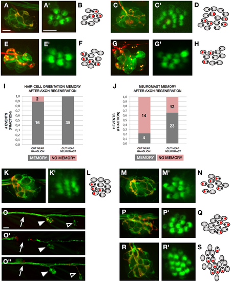
Regenerative synaptogenesis in neuromasts. (A–C) A neuromast of the transgenic line Tg[myo6b:actb1-EGFP; nkhgn39d] with mosaic expression of mCherry in a single LAN. (A) The individualized axon (red) synapses with crHCs. (A’) Magnified view of the hair bundles of hair cells in panel A. (B) Schematic representation of the example in panel A, in which hair cell orientation is indicated by an eccentric black dot and innervation by a red asterisk. (C) After severing, the individualized regenerated axon (red) recapitulate synapses with crHCs in the same neuromast. (C’) Magnified view of the hair bundles of hair cells in panel C. (D) Schematic representation of the example in panel C, indicating hair cell orientation (eccentric black dot) and synapses (red asterisk). (E) Neuromast double-transgenic Tg[myo6b:actb1-EGFP; Ribeye-Kusabira] showing a single LAN marked by mosaic expression of mCherry. The axon (red) synapses with rcHCs. (E’) Magnified view of the hair bundles of hair cells in panel E. (F) Schematic representation of the example in panel E, in which hair cell orientation is indicated by an eccentric black dot and innervation by a red asterisk. (G) After severing, the individualized axon (red) regenerated to recapitulate synapses with rcHCs. (G’) Magnified view of the hair bundles of hair cells in panel G. (H) Schematic representation of the example in panel G, indicating hair cell orientation (eccentric black dot) and synapses (red asterisk). (I) Quantification of hair cell innervation by regenerating singly marked axons of samples in which the axonal bundle was severed immediately below the neuromast (N = 35) (near neuromast) or furthest (N = 18) (near the posterior lateralis ganglion). All 35 axons cut near neuromast re-innervated hair cells of the original orientation—but 16 of the axons cut near the ganglion (0.89)—re-innervated the original orientation after regeneration (memory in grey), and 2 (0.11) did not (no memory in pink). (J) Quantification of neuromast innervation by regenerating singly marked axons of samples in which the lateralis nerves were severed immediately below the neuromast (near neuromast) (N = 35) or furthest (near the ganglion) (N = 18). A total of 23 (0.66) individualized axons cut near neuromast re-innervated the original neuromast (memory in grey), whereas 12 (0.34) re-innervated a different organ (no memory in pink). When axons were cut near the ganglion, 4 (0.22) re-innervated the original neuromast (memory in grey), and 14 (0.78) re-innervated a different organ (no memory in pink). (K–O”) Neuromast of the transgenic line Tg[myo6b:actb1-EGFP; nkhgn39d] with mosaic expression of mCherry in a single LAN. (K–K’) individualized axon (red) synapse with rcHCs. (K’) Magnified view of the hair bundles of hair cells in panel K. (L) Schematic representation of the example in panel K. (M) After axon severing and hair cell elimination, the individualized axon (red) regenerated to synapses with regenerated rcHCs in the same neuromast. (M’) Magnified view of the hair bundles of hair cells in panel M. (N) Schematic representation of the example in panel L–M. (O–O”) Selected time points from live confocal imaging of regenerative innervation of hair cells in the transgenic line Tg[myo6b:actb1-EGFP; nkhgn39d] (green) in an instance when a singly marked LAN (red) switched from a horizontal neuromast (solid arrowhead) to a vertical neuromast (empty arrowhead). Before laser-mediated severing (panel O), after severing (panel O’), and after regeneration (panel O”). The white arrow indicated the site of the cut. (P–P’, R–R’) Neuromast of the transgenic line Tg[myo6b:actb1-EGFP; nkhgn39d] with mosaic expression of mCherry in a single LAN. (P) The individualized axon (red) synapses with crHCs in a horizontal neuromast. (P’) Magnified view of the hair bundles of hair cells in panel P. (Q) Schematic representation of the example in panel P. (R) After severing, the individualized regenerated axon (red) switches to innervate a vertical neuromast, in which it synapses selectively with vdHCs. (R’) Magnified view of the hair bundles of hair cells in panel R. (S) Schematic representation of the example in panel R. Scale bars are 10 μm in A and A’ and 50 μm in O. crHC, caudorostral HC; HC, hair cell; LAN, lateral afferent neuron; rcHC, rostrocaudal HC; vdHC, ventrodorsal HC. |