
FIGURE
Fig. S2
- ID
- ZDB-FIG-180910-22
- Publication
- Heap et al., 2018 - Luminance Changes Drive Directional Startle through a Thalamic Pathway
- Other Figures
- All Figure Page
- Back to All Figure Page
Fig. S2
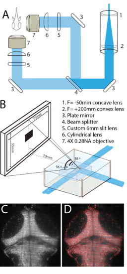
Optical setup for functional imaging during visual stimulation. (A) The selective plane illumination microscope (SPIM) setup used for all experiments. (B) Animals were placed in a custom-built chamber, embedded in agarose, with light from the SPIM entering from the front and side. A 75 X 55mm screen was placed 70mm from the animal for visual stimulation. (C) An example image of the tectum taken on the SPIM setup in (A), and the corresponding watershed segmentation of this image (D). |
Expression Data
Expression Detail
Antibody Labeling
Phenotype Data
Phenotype Detail
Acknowledgments
This image is the copyrighted work of the attributed author or publisher, and
ZFIN has permission only to display this image to its users.
Additional permissions should be obtained from the applicable author or publisher of the image.
Full text @ Neuron