
Fig. S1
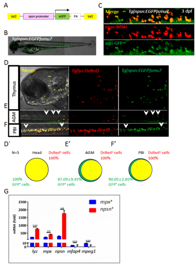
Generation and characterization of the Tg(npsn:EGFP)smu7 line. A. Construction of the Tg(npsn:EGFP)smu7 transgenic plasmid. A 2-kb upstream sequence of the zebrafish npsn gene (pink bar; referred to as the npsn promoter) was cloned and inserted prior to the GFP gene (green bar) and flanked by the two TP sites (yellow bar) in the Tol2 transgenic plasmid. B. The expression pattern of GFP signals in Tg(npsn:EGFP)smu7 at 3 dpf. C. Double fluorescent staining for GFP and npsn WISH in Tg(npsn:EGFP)smu7 at 3 dpf. (D–F) and (D’–F’). Inter-crossing of Tg(npsn:EGFP)smu7 and Tg(lyz:DsRed) lines to reveal overlapping populations. npsn+ cells overlapped 100% in the thymus (D and D’), 87.09 ± 9.45% in the AGM (E and E’), and 90.05 ± 2.85% in the PBI (F and F’) with mpx+ cells. White arrows indicate non-overlapping signals. G. The npsn promoter drives expression specifically in neutrophils. GFP+ cells sorted from Tg(npsn:EGFP)smu7 and Tg(mpx:EGFP). qRT-PCR showed neutrophil markers (lyz, mpx, and npsn) expressed at higher levels in npsn+ cells as compared with levels observed in mpx+ cells. The # represents “undetected”, [Mean ± SEM, n ≥ 200 in each group, triplicated], and statistical significance was determined using the two-tailed Student's t test. ***p < 0.001. |