
Fig. S3
- ID
- ZDB-FIG-180827-30
- Publication
- Wu et al., 2018 - A Rapid Method for Directed Gene Knockout for Screening in G0 Zebrafish
- Other Figures
- All Figure Page
- Back to All Figure Page
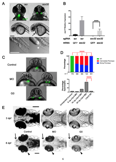
G0 Cas9 RNP Rescue and Comparison of G0 Cas9 RNP to Morpholino Knockdown. (Related to Figure 3) (A) Late sox32 phenotypes. Embryos were cell-injected at the one-cell stage with four-guide Cas9 RNP targeting sox32 or scrambled (scr) control, imaged at 24 hpf. Top panel showing Tg(myl7:GFP) labeling of the heart and cardia bifida after four-guide targeting of sox32. Middle panel showing cardiac edema (black arrow) after four-guide targeting of sox32. Bottom panel showing intestinal and cloacal agenesis (white asterisk). Scale bars 250 μm. (B) sox32 rescue. Embryos were yolk-injected at the one-cell stage with either four-guide Cas9 RNP targeting sox32 or scrambed control and either sox32 or GFP mRNA. Embryos were collected at 6 hpf and sox17 expression was measured by qPCR. **** p < 0.0001 one-way ANOVA with Tukey multiple comparisons test. Mean +/- SEM; n = 4 for each group. (C) Epifluorescence images of uninjected (Control), s1pr2 morpholino (MO), and s1pr2 four-guide Cas9 RNP-injected Tg(myl7:GFP) (G0) embryos at 48 hpf. Representative embryos are shown. About half of the MO-injected embryos showed incomplete fusion of the heart fields, an intermediate phenotype (left) with the remainder showing complete cardia bifida (right) but often less widely spaced than in four-guide Cas9 RNP-targeted embryos (bottom). Scale bar 250 μm. (D) Quantification of the phenotype penetrance and toxicity of embryos generated as in (A). Embryos were injected with MO or Cas9 RNP as indicated. Penetrance of the Strong s1pr2-specific phenotype was higher in Cas9 RNP G0 than in morphants at all MO doses tested. **** = p < 0.0001, Fisher’s exact test. The number of non-dysmorphic embryos scored is shown under each column. Percentage Dysmorphic increased with increasing s1pr2 MO dose (p < 0.0001; Chi-square test for trend). (E) Comparison of the durability of tyr Cas9 RNP (G0) or morpholino (MO) knockdown phenotypes. Representative embryos were analyzed at 3 and 8 dpf are shown. Arrows indicate areas of expected perioral pigmentation and arrowheads indicate retinal pigment epithelium at 8 dpf. Scale bars 250 μm. Note relatively similar levels of depigmentation in morphants and G0 Cas9 RNP embryos at 3 dpf but partial recovery of pigmentation in morphants by 8 dpf. Experiments were performed with yolk injections with doses denoted in Methods. All experiments were repeated at least once. |
Reprinted from Developmental Cell, 46, Wu, R.S., Lam, I.I., Clay, H., Duong, D.N., Deo, R.C., Coughlin, S.R., A Rapid Method for Directed Gene Knockout for Screening in G0 Zebrafish, 112-125.e4, Copyright (2018) with permission from Elsevier. Full text @ Dev. Cell