
Fig. 7
- ID
- ZDB-FIG-180827-27
- Publication
- Wu et al., 2018 - A Rapid Method for Directed Gene Knockout for Screening in G0 Zebrafish
- Other Figures
- All Figure Page
- Back to All Figure Page
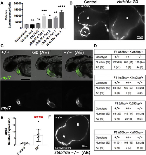
zbtb16a Disruption by Cas9 RNP Impairs Cardiac Development (A) Confirmation of increased nppb expression in response to zbtb16a disruption. Tg(nppb:F-Luc) embryos were yolk-injected with four distinct four-guide sets targeting zbtb16a or with control scrambled and slc24a5 guide sets, then collected at 2 dpf for luciferase assay. zbtb16a guide sets 1–3 targeted the coding sequence as in Figures 6A and 6B. zbtb16a guide set 4 specifically targeted the C2H2 zinc finger repeat domain. ∗p < 0.05, ∗∗p < 0.01, ∗∗∗p < 0.001, ∗∗∗∗p < 0.0001; ns, not significant compared with scrambled by one-way ANOVA and Sidak's multiple comparison test. Mean ± SEM; n = 24 for each group. (B) Representative spinning-disk confocal images of hearts from Tg(kdrl:GFP) embryos collected at 2 dpf after injection with Cas9 RNP with zbtb16a four-guide set (right) or a scrambled control set (left). Tg(kdrl:GFP) labels endocardium here. a, atrium; v, ventricle. (C) Representative images of zbtb16a wild-type Tg(myl7:GFP) embryo (left), Cas9 RNP-injected G0 embryo with atrial enlargement phenotype (AE) (middle), and F3 zbtb16a homozygous null embryo with AE phenotype (right) at 48 hpf. The AE phenotype includes marked chamber enlargement, pericardial edema, and impaired circulation. Fluorescent images at the bottom show myocardium. Scale bar, 250 μm. (D) Genotype and phenotype of F2 and F3 embryos derived from F1 zbtb16a Δ55bp heterozygous incrosses, F1 zbtb16a ins2bp heterozygous incross, F1 zbtb16a Δ7bp heterozygote/Δ55bp heterozygote transcross, and incross of F2 zbtb16a Δ55bp heterozygotes that had been outcrossed once with wild-type. All embryos were scored for AE phenotype at 2 dpf and then genotyped. (E) Quantification of nppb expression in AE phenotype embryos. AE phenotype embryos were selected from zbtb16a Δ55bp heterozygous incrosses and compared with clutchmate controls not exhibiting a cardiac phenotype. ∗∗∗∗ p < 0.0001, unpaired two-tailed t test. (F) Representative spinning-disk confocal image of heart from AE phenotype homozygous zbtb16a Δ55bp null Tg(myl7:GFP) embryo collected at 2 dpf. a, atrium; v, ventricle. |
Reprinted from Developmental Cell, 46, Wu, R.S., Lam, I.I., Clay, H., Duong, D.N., Deo, R.C., Coughlin, S.R., A Rapid Method for Directed Gene Knockout for Screening in G0 Zebrafish, 112-125.e4, Copyright (2018) with permission from Elsevier. Full text @ Dev. Cell