
Fig. S2
- ID
- ZDB-FIG-180827-29
- Publication
- Wu et al., 2018 - A Rapid Method for Directed Gene Knockout for Screening in G0 Zebrafish
- Other Figures
- All Figure Page
- Back to All Figure Page
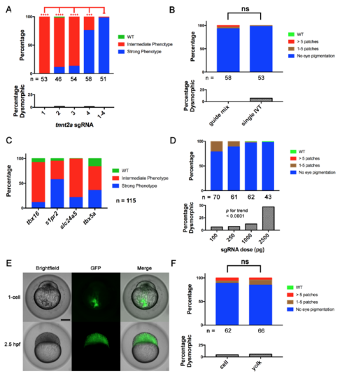
Four-Guide Set Cas9 RNP Generation, Dosing, and Route of Injection. (Related to Figure 2) (A) Penetrance of gene-specific phenotype and toxicity (as % dysmorphic) in embryos after single and four-guide Cas9 RNP injections targeting tnnt2a. **** = p < 0.0001, *** = p < 0.001 Fisher’s exact test for Strong vs. other phenotype (B) A four-guide Cas9 RNP mix for slc24a5 was generated by mixing individually produced sgRNAs (guide mix) or by pooled in vitro template elongation and transcription (single IVT). Cell-injection of either at the one-cell stage produced the slc24a5 depigmentation phenotype and toxicity at a similar rate at 2 dpf. (C) Cas9 RNP containing a pool of 4 sgRNAs targeting 4 different genes produced by a single template elongation and in vitro transcription was cell-injected into 1-cell stage embryos. Embryos were scored for the phenotypes of each individual gene disruption listed at 1 dpf (tbx16, s1pr2), 2 dpf (slc24a5), and 3 dpf (tbx5a). Control injections of a scrambled guide set demonstrated none of the phenotypes. Scoring was performed blinded to treatment. (D) % of embryos with depigmentation phenotype and % dysmorphic at 2 dpf after cell-injection of increasing doses of slc24a5 sgRNA preassembled with 800 pg of Cas9 protein. Increasing dose was associated with increasing toxicity. p < 0.0001 by Chi-square test for trend. (E) Transfer of scrambled sgRNA Cas9-GFP RNP to cells after yolk injection. Brightfield and epifluorescence images were acquired immediately and 2.5 hours after injection. Note complete transfer of fluorescence from injection site in yolk to cells within 2.5h. Scale bar 250 μm. (F) Comparison of yolk vs. cell injection of slc24a5 Cas9 RNP at the one-cell stage on pigmentation at 2 dpf. ns = non-significant for difference in rate of Strong phenotype or dysmorphic phenotype. All experiments were done at least twice with similar results. In A, B and D, the numbers of nondysmorphic embryos analyzed is shown below each column. |
Reprinted from Developmental Cell, 46, Wu, R.S., Lam, I.I., Clay, H., Duong, D.N., Deo, R.C., Coughlin, S.R., A Rapid Method for Directed Gene Knockout for Screening in G0 Zebrafish, 112-125.e4, Copyright (2018) with permission from Elsevier. Full text @ Dev. Cell