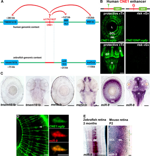
CNE1 enhancer activity is similar to miR-9 expression, but not mef2cb and tmem161b. (A) Schematic of the human and zebrafish genomic DNA containing CNE1 where SNP rs17421627 is located. (B) Confocal projections of EGFP immunolabeling in Tg(CNE1:egfp) containing the protective T allele or Tg(CNE1SNP:egfp) containing the risk G allele (rs17421627) in larvae at 72 hpf. In Tg(CNE1:egfp), EGFP expression is detected in cell bodies in the inner nuclear layer of the retina, telencephalon, optic tectum and hindbrain. The Tg(CNE1:egfp) expression pattern is reminiscent of miR-9 expression. EGFP expression in the CNS is abolished in the presence of the SNP rs17421627 risk allele. (C) Whole-mount ISH against tmem161b, mef2cb and miR-9 in larvae at 72 hpf. (D) Confocal section of double in situ/immunolabeling showing co-localization in the expression of endogenous miR-9 and EGFP protein in Tg(CNE1:egfp) retina at 72 hpf. (E) Whole-mount ISH against miR-9 in zebrafish (2 months old) or mouse (P2 stage) showing similar expression in the inner nuclear layer of the retina. Retina (R), Ganglion cells layer (GCL), Inner nuclear layer (INL), Telencephalon (T), Optic Tectum (OT), Hindbrain (H). Dorsal view of the brain with anterior up. Lateral view of the retina. Scale bars: 100 μm.
|
