
Fig. S1
- ID
- ZDB-FIG-180820-15
- Publication
- Madelaine et al., 2018 - A screen for deeply conserved non-coding GWAS SNPs uncovers a MIR-9-2 functional mutation associated to retinal vasculature defects in human
- Other Figures
- All Figure Page
- Back to All Figure Page
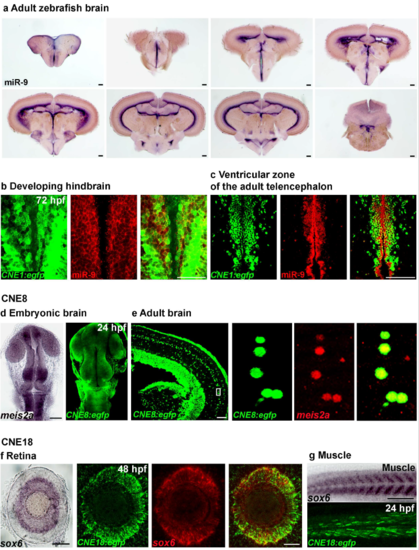
Functional validation of conserved CNEs enhancer activity and identification of the cis-regulated genes (a) Whole-mount in situ hybridization against miR-9 in the adult zebrafish brain. (b, c) Confocal section of double in situ/immunolabelling showing extensive overlap in the expression of endogenous miR-9 and EGFP protein in the hindbrain at 72 hpf (c) or in the ventricular zone of the adult zebrafish telencephalon (d) in the Tg(CNE1:egfp) line. (d) Tg(CNE8:egfp) expression is similar to meis2a endogenous expression at 24 hpf. (e) Confocal section of double in situ/immunolabelling showing the co-localization of endogenous meis2a and EGFP protein in the Tg(CNE8:egfp) adult brain. (f) Tg(CNE8:egfp) expression is similar to sox6 endogenous expression in the retina at 48 hpf. Confocal projection of double in situ/immunolabelling showing extensive overlap in the expression of endogenous sox6 gene and EGFP in Tg(CNE18:egfp) retina at 48 hpf. (g) Tg(CNE8:egfp) expression is similar to sox6 endogenous expression in muscle at 24 hpf. Dorsal view of the brain with anterior up. Lateral view of the retina. Lateral view of the trunk. Scale bars: 100 μm. |