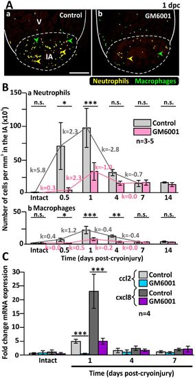
The numbers of inflammatory cells are reduced in the injured heart of GM6001-treated fish. (A) The Tg (coro1a:EGFP; lyz:dsRed) line of fish was used, where macrophages are labelled in green (see green arrowheads), and neutrophils are labelled in yellow (see yellow arrowheads). These images show the localization of macrophages and neutrophils in the injured areas (IA) of ventricles (V) at 1 dpc after the fish were injected with either (Aa) DMSO (controls) or (Ab) GM6001 (prepared in DMSO). The regions bounded by the white dashed lines indicate the IA, and scale bar: 200 µm. (B) Bar graphs to show the numbers of (Ba) neutrophils and (Bb) macrophages per mm3 in the IA of control and GM6001-treated fish at 0.5 dpc to 14 dpc, and in intact ventricles. The k values indicate the change in the numbers of cells per hour. (C) Bar graph to show the fold change in expression of cxcl8 and ccl2 in the intact heart and at 1 dpc, 4 dpc and 7 dpc in both control and GM6001-treated fish. In (B) and (C), the data are expressed as the mean ± standard deviation of n = 3–5 and n = 4 experiments, respectively, and the asterisks indicate GM6001 data that are significantly different from the DMSO controls at p < 0.05 (*), p < 0.01 (**) and p < 0.001 (***); one-way ANOVA with LSD Post-hoc test.
|
