
Fig. 1
- ID
- ZDB-FIG-180706-7
- Publication
- Siddam et al., 2018 - The RNA-binding protein Celf1 post-transcriptionally regulates p27Kip1 and Dnase2b to control fiber cell nuclear degradation in lens development
- Other Figures
- All Figure Page
- Back to All Figure Page
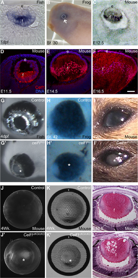
Celf1 is required for vertebrate lens development. (A) In zebrafish, celf1 transcripts are detected in the lens at 1 day post fertilization (1dpf) by in situ hybridization (ISH). (B) In X. laevis, ISH indicates strong celf1 expression (arrow) in the embryonic St. 30 eye (arrow) and lens (indicated by broken line area). (C) In mouse, ISH shows strong Celf1 transcript expression in the lens at embryonic day 12.5. (D) In mouse lens, Celf1 protein is expressed at (D) E11.5 (E) E14.5 and (F) E16.5 predominantly in fiber cells (f) and to a lower extent in epithelial cells (e). (G, G’) In zebrafish, while control eyes are normal, celf1 knockdown (KD) results in microphthalmia and clouding of lens (asterisk) by 4dpf. (H, H’) In X. laevis, compared to control, celf1 KD results in microphthalmia. (I, I’) In mouse, compared to control, Celf1cKO/cKO lens exhibits severe cataract (asterisk). (J-K’) Compared to control, refraction errors (asterisk) are observed in Celf1cKO/cKO lens under dark-field and light-field microscopy. (L, L’) At E16.5 stage, the mouse Celf1cKO/cKO lens exhibits abnormal spaces (asterisk) in the fiber cell region. Scale bar in F is 75 μm. |
| Gene: | |
|---|---|
| Fish: | |
| Anatomical Term: | |
| Stage: | Prim-5 |
| Fish: | |
|---|---|
| Knockdown Reagent: | |
| Observed In: | |
| Stage: | Day 4 |