
Fig. S9
- ID
- ZDB-FIG-180706-13
- Publication
- Siddam et al., 2018 - The RNA-binding protein Celf1 post-transcriptionally regulates p27Kip1 and Dnase2b to control fiber cell nuclear degradation in lens development
- Other Figures
- All Figure Page
- Back to All Figure Page
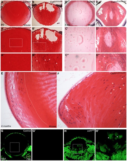
Celf1 deficiency-mediated nuclear degradation defects are persistent. (A to B”) In mice, compared to control, Celf1cKO/cKO lens at stage P0 exhibits fiber cell nuclear degradation defects. Note the abnormal presence of nuclei (asterisks) in the central region of the fiber cells. A’ to B” are higher magnification images of A and B, respectively. (C to D”) Compared to control, Celf1lacZKI/lacZKI lens at seven-week age continue to exhibit nuclear degradation defects. Note the abnormal presence of nuclei (asterisks) in the central fiber cell region. C’ to D” are higher magnification images of C and D, respectively. (E and F) Compared to control where a clear nuclear free zone is visible (broken white line), Celf1lacZKI/lacZKI mouse lens at 4 months continue to exhibit nuclear degradation defects. Note the abnormal presence of nuclei (asterisks) in the central fiber cell region. (G to H’) Compared to control, zebrafish celf1 KD lens exhibits nuclear degradation defects at stage 5dpf. Note the abnormal presence of nuclei (asterisks) in the central fiber cell region. E’ and F’ are high-magnification of the dotted-line area in E and F. |
| Fish: | |
|---|---|
| Knockdown Reagent: | |
| Observed In: | |
| Stage: | Day 5 |