
Fig. S1
- ID
- ZDB-FIG-180706-10
- Publication
- Siddam et al., 2018 - The RNA-binding protein Celf1 post-transcriptionally regulates p27Kip1 and Dnase2b to control fiber cell nuclear degradation in lens development
- Other Figures
- All Figure Page
- Back to All Figure Page
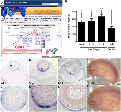
Celf1 mRNA expression in vertebrate lens development. (A)iSyTE identifies Celf1 as a highly lens-enriched gene in lens development. Lens enrichment extent is indicated by differing red color intensities in the heat-map. (B) Analysis of iSyTE lens microarray datasets from mouse embryonic stages 10.5, 11.5 and 12.5 shows highly enriched expression of Celf1 compared to whole embryonic body tissue (WB) control. Celf1 microarray probe binding fluorescent signal intensity is represented on the Y-axis and the different mouse embryonic lens stages are shown on the X-axis. (C) At 2dpf (day post fertilization), (D) 3dpf and (E) 4dpf, zebrafish lenses exhibit expression of celf1 mRNA in the transition zone (arrow in C) and in later stages in the posterior region (arrows in D and E). (C’ to E’) High-magnification of C to E. (F) In Xenopus laevis, celf1 mRNA expression is observed from early developmental stage (St. 23) in the eye region (arrow; lens area indicated by broken white line). (F’) High-magnification of F. Lens area is indicated by broken white line. Asterisks in B represents a p-value less than 0.05. |
| Gene: | |
|---|---|
| Fish: | |
| Anatomical Term: | |
| Stage Range: | Long-pec to Day 4 |