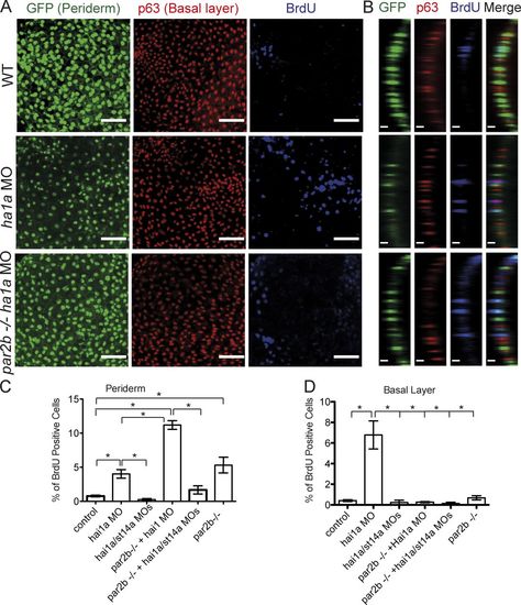
Effect of Par2b deficiency on increased BrdU incorporation associated with loss of hai1a function in the basal layer versus periderm. (A and B) Control and hai1a morphants periderm-nuclear-EGFP embryos with and without St14a or Par2b deficiency were incubated with BrdU for 3 h beginning at 28 hpf, collected for analysis at 46 hpf, and immunostained for p63 and BrdU. Bar, 50 µm. (B) Colocation of BrdU staining with either nuclear-GFP or nuclear p63 staining was used to identify periderm or basal layer cells that had incorporated BrdU, respectively. (C and D) The percentage of BrdU-positive nuclei in periderm (C) and basal layer (D) is shown (mean ± SEM of three experiments; for each experiment, 10 embryos were examined per condition, and 150–200 cells were analyzed in each embryo). Results were analyzed by one-way ANOVA and Bonferroni posttest. In C, the hai1a/st14a MO group and the par2b−/− hai1a/st14a MO groups were not different from control; the hai1a MO group, the par2b−/− group, and the par2b−/− hai1a MO group were all different from control, and the par2b−/− and hai1a MO groups were different from the par2b−/− hai1a MO group (*, P < 0.05). In D, the hai1a MO group was different from all other groups (*, P < 0.0001).
|
