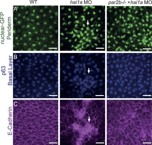
Altered distribution of nuclei and E-cadherin in basal-layer keratinocytes in hai1a morphants and its par2b-dependence. Control, hai1a morphant, and par2b−/− hai1a morphant periderm-nuclear-GFP embryos were collected at 30 hpf, immunostained for the basal layer marker p63 and E-cadherin, and imaged by confocal fluorescence microscopy. (A) Nuclear-GFP marker of periderm cells. Note regular spacing in controls (left), clustered and crowded appearance in hai1a morphants (middle), and increased number in par2b−/− hai1a morphants (right). (B) p63 marker for basal layer nuclei. Note regular spacing in controls (left), clustered and crowded appearance in hai1a morphants (middle), and more control-like pattern in par2b−/− hai1a morphants (right). (C) E-cadherin staining in basal layer. Note localization to cell–cell junctions in controls (left), aberrant distribution with increased cytosolic staining in areas coinciding with clusters of p63-positive nuclei in hai1a morphants (middle), and more control-like pattern in par2b−/− hai1a morphants (right). Bar, 20 µm.
|
