
FIGURE
Fig. 2
- ID
- ZDB-FIG-180620-68
- Publication
- Minegishi et al., 2018 - Mutation in the Zebrafish cct2 Gene Leads to Abnormalities of Cell Cycle and Cell Death in the Retina: A Model of CCT2-Related Leber Congenital Amaurosis
- Other Figures
- All Figure Page
- Back to All Figure Page
Fig. 2
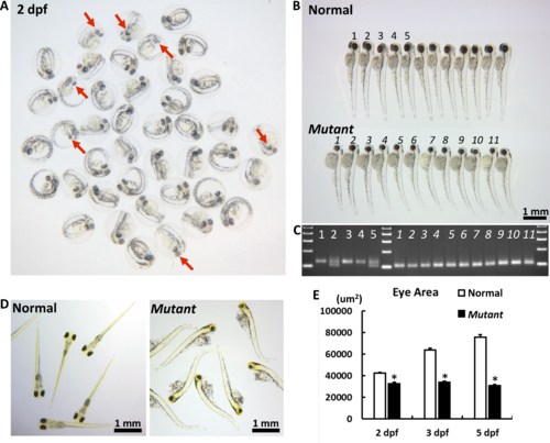
The macroscopic eye phenotype of the cct2-L394H-7del mutant. (A) The representative picture of F3 embryos from F2 cross mating at 2 dpf. At this stage, some embryos had smaller eyes with reduced pigmentation (marked by red arrows) compared with most embryos. (B) The representative picture of F3 embryos from F2 cross mating at 3 dpf. At this stage, the homozygous cct2-L394H-7del mutant embryos (lower row) were easily distinguished from wild-type and heterozygous cct2-L394H-7del embryos (upper row) by smaller size and circular shape of their eyes. The numbering (1–5 for normal eye and 1–11 for smaller eye larvae) is identical to the numbering in (C). (C) The genotyping of embryos labeled in (B) by PCR. The wild-type allele produces a 125-bp band, while the mutant allele produces a 101-bp band. (D) The representative picture of F3 embryos from F2 crossmating at 5 dpf. At this stage, wild-type and heterozygous larvae start to swim, while homozygous mutant larvae are not able to swim. (E) Comparison of the eye area between normal versus homozygous cct2-L394H-7del mutant embryos at 2, 3, and 5 dpf. *P < 0.05.
|
Expression Data
Expression Detail
Antibody Labeling
Phenotype Data
| Fish: | |
|---|---|
| Observed In: | |
| Stage Range: | Long-pec to Day 5 |
Phenotype Detail
Acknowledgments
This image is the copyrighted work of the attributed author or publisher, and
ZFIN has permission only to display this image to its users.
Additional permissions should be obtained from the applicable author or publisher of the image.
Full text @ Invest. Ophthalmol. Vis. Sci.