
FIGURE SUMMARY
- Title
-
Mutation in the Zebrafish cct2 Gene Leads to Abnormalities of Cell Cycle and Cell Death in the Retina: A Model of CCT2-Related Leber Congenital Amaurosis
- Authors
- Minegishi, Y., Nakaya, N., Tomarev, S.I.
- Source
- Full text @ Invest. Ophthalmol. Vis. Sci.
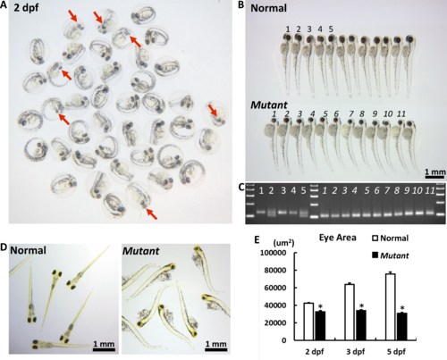
The macroscopic eye phenotype of the cct2-L394H-7del mutant. (A) The representative picture of F3 embryos from F2 cross mating at 2 dpf. At this stage, some embryos had smaller eyes with reduced pigmentation (marked by red arrows) compared with most embryos. (B) The representative picture of F3 embryos from F2 cross mating at 3 dpf. At this stage, the homozygous cct2-L394H-7del mutant embryos (lower row) were easily distinguished from wild-type and heterozygous cct2-L394H-7del embryos (upper row) by smaller size and circular shape of their eyes. The numbering (1–5 for normal eye and 1–11 for smaller eye larvae) is identical to the numbering in (C). (C) The genotyping of embryos labeled in (B) by PCR. The wild-type allele produces a 125-bp band, while the mutant allele produces a 101-bp band. (D) The representative picture of F3 embryos from F2 crossmating at 5 dpf. At this stage, wild-type and heterozygous larvae start to swim, while homozygous mutant larvae are not able to swim. (E) Comparison of the eye area between normal versus homozygous cct2-L394H-7del mutant embryos at 2, 3, and 5 dpf. *P < 0.05.
|
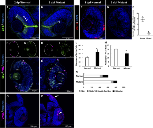
The retinal phenotype of the homozygous cct2-L394H-7del mutants at 2 pdf. (A, B) Staining of wild-type (A) and homozygous cct2-L394H-7del mutant (B) retinas with antibodies against Zn-5. Note a dramatic reduction of Zn-5 staining of the retinal ganglion cell layer (marked by a white bracket) and thinner optic nerve in cct2-L394H-7del mutant compared with wild-type. The lamination of the neural retina was more pronounced in wild-type embryos than in mutants. (C, D) The cone photoreceptor was underdeveloped in the homozygous cct2-L394H-7del mutants at 3 dpf. Staining of wild-type (C) and homozygous cct2-L394H-7del mutant (D) retinas with antibodies against zpr-1. (E) Quantification of zpr-1–positive cells in wild-type and homozygous cct2-L394H-7del mutant retinas (n = 4). (F–I) The distribution of PH3-positive and EdU-positive cells in the retina. In wild-type embryos, PH3-positive cells (F) and most of EdU-positive cells (G) were found in the ciliary marginal zone and the area of neuronal precursors localization. In the retina of homozygous cct2-L394H-7del mutants, PH3-positive cells were found in the ciliary marginal zone, area of neuronal precursors localization and lens epithelium (H). EdU-positive cells were found through the neural retina, in the ciliary marginal zone and lens epithelium (I). The white dashed lines indicate the eyecup boundaries. (J–K) Merged images of EdU and PH3 staining of the retina of wild-type (J) and homozygous cct2-L394H-7del mutant (K). (L–N) Quantification of EdU-positive, PH3-positive, and EdU/PH3-double positive cells in wild-type and homozygous cct2-L394H-7del mutant retinas (n = 10). (O, P) TUNEL staining of wild-type (O) and mutant (P) retinas. Arrow marks the optic tectum, the area of retinal ganglion cell axon projection; arrowhead marks TUNEL-positive cells in the neural retina. CMZ, ciliary marginal zone; Ln, lens. Scale bars: 50 μm. *P < 0.05.
EXPRESSION / LABELING:
PHENOTYPE:
|

Rescue of a small eye phenotype by CCT2 RNA injection. (A) Representative pictures of 2 dpf embryos after injection of wild-type human CCT2 RNA (100 pg). Genotyping of injected embryos is shown below. Ref. panel shows uninjected cct2-L394H-7del mutant. (B) Western blot analysis of indicated proteins in 2-dpf embryos after injection of RNA encoding wild-type or LCA-causing T400P CCTβ. Normal and mutant lines correspond to uninjected control and mutant larvae. Numbers below each lane indicate changes in the protein level relatively to uninjected normal larvae after normalization using Poncer S (Pon. S) staining. Two independent sets of embryos were analyzed. (C) Western blot analysis of indicated proteins in 3-dpf embryos. Three independent sets of embryos were analyzed. (D–E) The zn-5 staining of homozygous 3-dpf cct2-L394H-7del mutant eye section after mocked (D) or wild-type CCT2 RNA (E) injection. (F) The zn-5 staining of 3-dpf wild-type eye section after wild-type CCT2 RNA injection. Size-rescued mutant retinas demonstrated zn-5 staining and lamination comparable to those of wild-type retina. (G–I) The TUNEL staining of the eye sections as in (D–F). Note the absence of TUNEL-positive cells in the size-rescued mutant retina.
|
Acknowledgments
This image is the copyrighted work of the attributed author or publisher, and
ZFIN has permission only to display this image to its users.
Additional permissions should be obtained from the applicable author or publisher of the image.
Full text @ Invest. Ophthalmol. Vis. Sci.