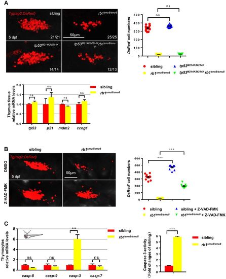
rb1 deficiency induced apoptosis in a caspase-dependent manner. (A) Confocal images of rag2:DsRed cells in the thymus of the siblings, rb1smu8/smu8 mutants, tp53M214K/M214K and tp53M214K/M214Krb1smu8/smu8 double mutants at 5 dpf. Scale bars: 50 μm. Quantification of DsRed+ cell numbers in the thymus (right). Expression of tp53, p21, mdm2 and ccng1 in thymocytes excised from siblings and rb1-deficient embryos at 5 dpf (bottom) (mean±s.e.m.; ns, not significant; n=11). (B) Confocal images of rag2:DsRed cells in the thymus of DMSO-treated siblings, DMSO-treated rb1smu8/smu8 mutants, Z-VAD-FMK-treated siblings and Z-VAD-FMK-treated rb1smu8/smu8 mutants at 5 dpf. Scale bars: 50 μm. Quantification of DsRed+ cell numbers in the thymus of sibling embryos and rb1smu8/smu8 mutants (right) (mean±s.e.m.; ***P<0.001; n=10). (C) Expression of caspase mRNA in thymocytes excised from siblings and rb1-deficient embryos at 5 dpf (mean±s.e.m.; *P<0.05; ***P<0.001; ns, not significant; n=30). The activity of caspase 3 in siblings and rb1smu8/smu8 embryos is expressed as the fold change compared with siblings (mean±s.e.m.; ns, not significant; n=10).
|
