
Fig. S3
- ID
- ZDB-FIG-180509-10
- Publication
- Meier et al., 2017 - Cohesin facilitates zygotic genome activation in zebrafish
- Other Figures
- All Figure Page
- Back to All Figure Page
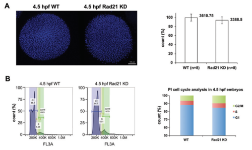
Cell cycle analysis of Rad21-depleted embryos (A) Left, whole mount wild type (WT) and Rad21-depleted (Rad21 KD) embryos at 4.5 hpf. Embryos were fixed in 4% formaldehyde, dechorionated, and dehydrated in methanol. Nuclei were stained with hoechst, and confocal Z-stacks of embryos were obtained (see methods). Right, nuclei were quantified using lmaris software. No significant difference was observed in nuclei numbers between Rad21 KD and WT (n=8, p=0.1138, un-paired t-test). (B) Cell cycle analysis of WT and Rad21 KD embryos. Let, representative propidium iodide staining profiles of 4.5 hpf WT and Rad21 KD embryos, with G1, S, G2/M phases indicated. Right, Rad21 KD embryos had ~50% more cells in G2/M phase compared to WT controls. |