
Fig. S1
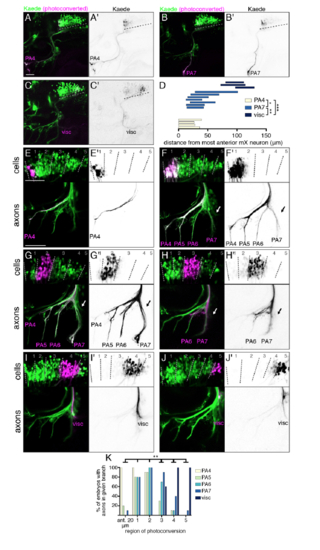
Topographic mapping by vagus motor neurons. Related to Figure 1. (A-C) Photoconverted Kaede in a single one of the mX axon branches at 4 dpf is shown in magenta and (A’-C’) black. Dotted lines mark the length of the mX territory. PA4-innervating neurons are at the anterior edge of the mX territory, PA7-innverating neurons are posterior to PA4 neurons, and viscera-innervating neurons are at the posterior-most end of the mX territory. Unfortunately, due to the small scale of the region, we were not able to photoconvert PA5 or PA6 alone. (D) Quantification of axon photoconversion experiments. Each bar represents a single embryo. Analysis done using unpaired t test comparing anterior and posterior borders of photoconverted population. (E-J) Photoconverted Kaede in cell bodies is shown in magenta and (E’-J’) black with anterior left and dorsal up. Bottom panels show photoconverted Kaede in peripheral axon branches. (E,E’) The anterior-most 20 um of the mX territory was photoconverted. (F-J,F’-J’) 5 regions were defined using somite boundaries, with the anterior border of region 1 aligning with the anterior border of somite 1, and the posterior border of region 5 extending halfway into somite 2. Cells in a single region were photoconverted. (K) Quantification of (E-J). n = 10 embryos per region photoconverted. Analysis was done using Chi-square test comparing regions in pairwise combinations with respect to each axon branch. Regions are considered different if they differ in at least one branch. Anterior left and dorsal up, scale bars 50 um. |