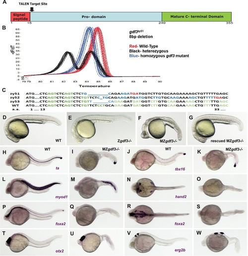
(A–C) gdf3 mutants were generated using TALEN-mediated mutagenesis. (A) Gdf3, a TGFβ family member, comprises a signal peptide, pro-domain and mature TGFβ domain. TALENs were designed to target genomic sequences located near the amino end of the pro-domain. (B) Mutants were identified by high-resolution melt analysis (HRMA). (C) Three mutant alleles, gdf3zy51, gdf3zy52, gdf3z53, had 8, 1 and 6 bp deletions respectively. (D–G) Morphological phenotypes of gdf3 mutants at 24 hpf. (D) Wild-type (WT) and (E) zygotic (Z) gdf3 mutants were phenotypically indistinguishable. (F) Maternal-zygotic (MZ) gdf3 mutants lacked notochord, pharyngeal endoderm and had reduced anterior neural tissues. (G) MZgdf3 mutants were completely rescued by injection of 100 pg gdf3 RNA at the one-cell stage. (H–W) WISH analysis of gene expression in WT (columns H-T and J-V) and MZgdf3 (columns I-U and K-W) mutants at 24 hpf. (H, I) ta (ntl) expression in the notochord was absent from MZgdf3 although tailbud expression was maintained. (J, K) tbx16 (spt) expression in spinal cord neurons was absent in MZgdf3 while tailbud expression is unaffected. (L, M) Trunk and tail somites expressing myod1 were reduced in number and altered in shape in MZgdf3. (N, O) Expression of hand2 in the heart, pharyngeal arch mesoderm and pectoral fin buds is absent in MZgdf3. (P–S) Ventral neural tissues and pharyngeal endoderm expressing foxa2 (axial) were absent in MZgdf3. Patterns of expression of otx2 in the forebrain and midbrain (T, U), and erg2b (krox20) in hindbrain rhobomeres 3 and 5 (V, W) were altered in MZgdf3 mutants. All embryos (D–W) are shown in lateral view with rostral to the left except N, O, R, S which are dorsal views in the same orientation. Each panel is a representative image from at least 15 embryos.