|
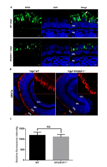
Immunostaining of retinal sections of wildtype and rpgrip1-/- zebrafish using anti-RPGR antibody showed abnormal localization of Rpgr. Rpgr was localized to the connecting cilia and outer segments of photoreceptors in 14dpf wildtype zebrafish retina, while Rpgr was mislocalized in inner segments of photoreceptor cells of rpgrip1-/- mutant retinas and the fluorescence signals were significantly decreased. Arrow shows rod outer segment. (B) There is no difference in GRK7a localization to the cone outer segments in both wildtype and rpgrip1 mutant zebrafish at 5dpf. (C)The fluorescence signals of the boxed areas of wildtype and mutant retinal sections were measured using Image J software. There is no significant difference between wildtyep and mutants with p=0.6989. GCL, ganglion cell layer; INL, inner nuclear layer; ONL, outer nuclear layer; RPE, retinal pigment epithelium. NS, no significance.
|