FIGURE

Fig. 3

ID
ZDB-FIG-180322-19
Publication
Wang et al., 2017 - The construction of intrahepatic cholangiocarcinoma model in zebrafish
Other Figures
All Figure Page
Back to All Figure Page
Fig. 3

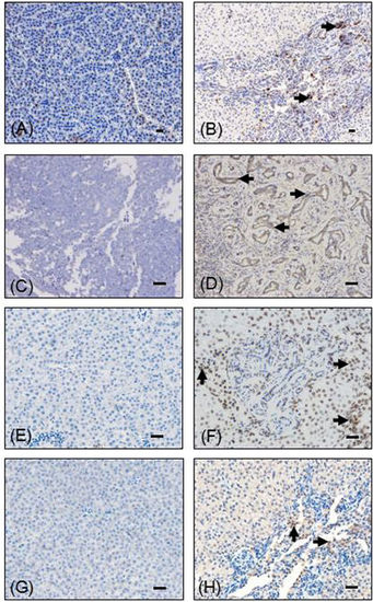
Immunohistochemical examination of ICC in WT and Tg(fabp10:nras 61K) transgenic zebrafish at 12 mpf. Immunohistochemical images of PCNA (A), CK19 (C), phosphorylated ERK (pERK) (E), phosphorylated MEK1/2 (pMEK1/2) (G) in WT and Tg(fabp10:nras 61K) transgenic zebrafish at 12 mpf. Tumor surrounding tissue of Tg(fabp10:nras 61K) transgenic zebrafish liver showing intense PCNA (B), CK19 (D), pERK (F), pMEK1/2 (H) immunostaining in Tg(fabp10:nras 61K) transgenic zebrafish at 12 mpf. Immunostaining signals in the bile duct of Tg(fabp10:nras 61K) transgenic zebrafish liver are indicated by arrowheads. Scale bars: 50 µm.

Expression Data
Antibodies:
Fish:
Anatomical Terms:
Stage: Adult

Expression Detail
Antibody Labeling
Phenotype Data
Fish:
Observed In:
Stage: Adult

Phenotype Detail
Acknowledgments
This image is the copyrighted work of the attributed author or publisher, and ZFIN has permission only to display this image to its users. Additional permissions should be obtained from the applicable author or publisher of the image. Full text @ Sci. Rep.