|
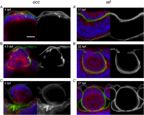
Disruption of basement membrane (capsule) integrity in the mutant occ lens. When the epithelial cells of the occ lens became multilayered at 4 dpf, the basement membrane appeared as a thin disorganized layer on the external surface of the lens mass. By 4–5 dpf (A–C), a thin capsule was developing to surround the occ lens and appeared to be distributed irregularly around the lens cell mass. Apparently, the discontinuities in the basement membrane destabilized the structure enough to permit epithelial cells to form a secondary cell mass by 5 dpf. In contrast, the basement membrane of the wt lens formed a thick, prominent, and continuous capsule surrounding the lens mass as early 22 hpf (A′,B′), indicating the earliest stages of lens capsule development. Sections were labeled for laminin (green), actin (red), and DAPI (blue) and shown with corresponding monochrome images (to the right) of the green laminin channel only. Approximate ages are listed in each section. Scale bar = 20 μm.
|