
Fig. 5
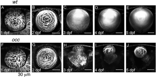
Lens Development in occ crossed with Q01. The 2-photon live-embryo imaging of lenses from wt and occ crossed to Q01 was conducted at 1, 2, 3, 4, and 5 dpf. The cornea is at the top of each image and the retina is at the bottom. The lens development in occ was the same as in the wt until 2– 3 dpf, when the lenticular cell mass separated from the surface ectoderm and the lens epithelial cells differentiated into secondary lens fibers. After 3 dpf, a nucleus began to form in the center of the wt lens, and the lens grew larger gradually and matured similar to an adult lens. In contrast, the lens in the occ eye was abnormal. Epithelial cells formed a second lenticular cell mass on the anterior side of the first lens, and the first lens began degenerating at 3 dpf. At 4–5 dpf, the secondary lens cell mass resembled a normal 2-dpf lens. See Suppl. Movies 1–3. Scale bar = 30 μm. |
| Fish: | |
|---|---|
| Observed In: | |
| Stage Range: | Prim-5 to Day 5 |