
Fig. S7
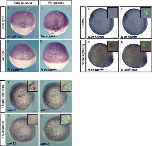
N-cadherin is expressed downstream of Nodal but does not induce an endodermal fate. (A–D) N-cadherin expression detected by in situ hybridization is lost in the shield and hypoblast in mutants lacking Nodal signaling (MZoep mutants). dfc, dorsal forerunner cells; hyp, hypoblast; sh, shield. (E–H) Conversely, N-cadherin expression is induced by Nodal signaling. Control ectodermal cells (E and F) or cells expressing the constitutively active Nodal receptor (G and H) were transplanted to the animal pole of a host embryo. N-cadherin expression was revealed by in situ hybridization. E–H show the same embryos in brightfield alone (E and G) or with the green signal from transplanted cells (F and H). Insets are close-ups of the animal pole region bearing transplanted cells (magnification, 3×). (I–L) Contrary to activation of Nodal signaling (I and J), which was used as a control, overexpression of N-cadherin (K and L) does not induce an endodermal fate (in situ hybridization for sox32) in cells transplanted to the animal pole. I–L show the same embryos in brightfield alone (I and K) or with the green signal from transplanted cells (J and L). Insets are close-ups of the animal pole region bearing transplanted cells (magnification, 3×). (Scale bars: 100 μm.) |
| Gene: | |
|---|---|
| Fish: | |
| Anatomical Terms: | |
| Stage Range: | 50%-epiboly to 75%-epiboly |