
Fig. S2
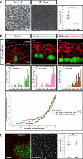
Endodermal cell internalization is independent of E-cadherin levels. (A) E-cadherin expression, assessed by immunohistochemistry, was reduced upon morpholino injection (animal views). Images were all acquired in the same conditions. (Scale bar: 20 µm.) Fluorescence intensity was quantified at cell boundaries (n = 6 embryos for each condition). ***P < 0.001. (B) To assess the importance of E-cadherin adhesion in internalization, endodermal cells were transplanted to the animal pole, E-cadherin being down-regulated either in transplanted endodermal cells or in their ectodermal neighbors. (Top) Sagittal sections showing the position of transplanted cells at midgastrulation. (Scale bar: 10 µm.) (Middle and Bottom) Distribution of embryos according to the percentage of internalized cells at midgastrulation plotted as histograms (Middle) or as a cumulative plot (Bottom). (C) E-cadherin expression assessed by immunohistochemistry in induced endodermal cells (expressing GFP) and ectodermal cells (animal view). Fluorescence intensity was quantified at boundaries between ectodermal cells and between endodermal cells (n = 5 embryos, P = 0.6). (Scale bar: 20 µm.) ns, nonsignificant (P > 0.05). |