
Fig. 7
- ID
- ZDB-FIG-180118-27
- Publication
- Bayramli et al., 2017 - Patterned Arrangements of Olfactory Receptor Gene Expression in Zebrafish are Established by Radial Movement of Specified Olfactory Sensory Neurons
- Other Figures
- All Figure Page
- Back to All Figure Page
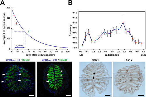
Cell death in the zebrafish OE. (A) Immunohistochemistry against BrdU and HuC/D and survival curve of BrdU/HuC double-positive OSNs (top; mean ± SEM; top) following 24 h incubation with BrdU. The data points after the initial increase of double positive cells between 2d and 4d can be fitted with the exponential function #cells = 387.5e−0.035 X, which corresponds to an OSN half life of t1/2 = 19.8 d. (n = 2d: 17, 4d: 26, 8d: 18, 30d: 51, and 90d: 60 sections). Representative cross sections for the 4d (left) and 90d (right) time point are shown (bottom). BrdU/HuC double-positive cells (turquoise, arrowheads) are more numerous at earlier time points and undergo positional shifts towards the centre of the sensory OE. (B) Detection of cell death in the adult zebrafish OE by terminal deoxynucleotidyl transferase dUTP nick end labeling (TUNEL) of fragmented DNA on cross sections though the OEs of two different zebrafish (bottom). TUNEL-positive cells appear dark brown by chromogenic detection with 3,3′-diaminobenzidine. The images represent composites of two high-resolution pictures joined together at indicated positions (dotted lines) to cover the full sections. The frequency distribution of TUNEL-positive cells along the radial dimension of the sensory region of the OE is shown in the top graph. The position of cells has been normalized to the distance of the sensory/nonsensory border (SNS) from the tip of the interlamellar curve (ILC). TUNEL-positive cells can be found throughout the entire sensory OE but are unevenly distributed and more abundant in the vicinity of the ILC and in the centre of the sensory OE. The data points represent the means ± SEM of the distribution of 689 cells from 9 sections of 3 fish. Scale bars: 100 µm. |