
Fig. 6
- ID
- ZDB-FIG-180118-26
- Publication
- Bayramli et al., 2017 - Patterned Arrangements of Olfactory Receptor Gene Expression in Zebrafish are Established by Radial Movement of Specified Olfactory Sensory Neurons
- Other Figures
- All Figure Page
- Back to All Figure Page
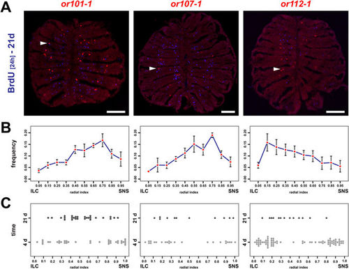
OSNs continue to undergo positional shifts after onset of OR expression. (A) Immunohistochemistry against BrdU (blue) on cross sections through the OE of adult zebrafish that were incubated for 24 h in BrdU 21d prior to analysis and simultaneous in situ-hybridization (red) for the three olfactory receptors or101-1 (left), or107-1 (centre), and or112-1 (right). For each receptor BrdU double-positive cells (arrowheads) can be identified. Scale bar: 100 µm. (B) OR frequency distribution profiles across the radial dimension of the OE for the receptors shown in A (left: or101-1, centre: or107-1, right: or112-1). The position of each cell was normalized to the distance of the sensory/nonsensory border (SNS) from the tips of the interlamellar curves (ILCs). The data represent the positions (mean ± SEM) of: or101-1: 755 cells from 4 OEs of 4 fish, or107-1: 332 cells from 4 OEs of 4 fish, and or112-1: 464 cells from 6 OEs of 3 fish. (C) Spatial distribution of young (4d, light grey) and old (21d, dark grey) OSNs expressing or101-1 (left), or 107-1 (centre), and or112-1 (right) normalized to the length of the sensory OE between the tips of the interlamellar curves (ILCs) and the sensory/nonsensory border (SNS). OSNs expressing specific OR genes continue to undergo positional shifts over time. |
| Genes: | |
|---|---|
| Fish: | |
| Anatomical Term: | |
| Stage: | Adult |