|
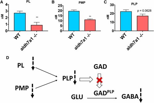
B6 vitamers are changed in aldh7a1−/− larvae compared to WT siblings. Lower levels of PL (A), PMP (B), and PLP (C) were observed in the null-mutants compared to WT according to liquid chromatography-mass spectrometry analysis using polar metabolite extracts (three replicates of six larvae pools). Asterisks indicate statistical significance according to Student’s t-test (* P < 0.05 and ** P < 0.01). Error bars represent ± SD. Possible mechanism for low GABA levels observed in the mutant larvae correlating with lower B6 vitamer levels (D), with potential reduction in the conversion levels of the inactive apo-form of GAD (and other PLP-dependent enzymes) to their catalytically active holo-form (GADPLP) by the covalent attachment of PLP. GABA, γ-aminobutyric acid; GAD, glutamate decarboxylase; PL, pyridoxal; PMP, pyridoxamine 5′-phosphate; PLP, pyridoxal 5′-phosphate; WT, wild-type.
|