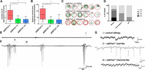
Seizure phenotype in aldh7a1−/− 11-dpf larvae. Video recordings (n = 12 larvae per genotype) were analyzed for high-speed movement in the presence of light stimulus; the calculated duration (A) and distance traveled (B) are shown. Movement traces shown in red, green, and black represent high-speed, intermediate, and slow movements, respectively (C). Blinded classification of seizure scores from video recordings of mutants, heterozygous, and WT larvae (n = 12 larvae per genotype). S0 characterizes normal swimming behavior, S1 reflects increased activity, S2 indicates rapid circular swimming activity, and S3 represents whole-body convulsions. Tectal field recordings of representative 11-dpf WT sibling (E) and aldh7a1−/− (F) showing spontaneous epileptiform-like electrographic activity in the mutants. Amplified view (G) of regions of the traces shown in (E and F): “i” normal activity as seen in WT and heterozygous siblings, “ii” ictal-like, and “iii” interictal-like epileptiform discharge observed in the mutants. Asterisks indicate statistical significance (* P < 0.05, ** P < 0.01, and *** P < 0.001) according to one-way ANOVA test on top of the graphics (in (A) P = 0.0003 and in (B) P = 0.0002) with Tukey’s post hoc pairwise tests. Error bars represent ± SD. All experiments were performed comparing aldh7a1−/− and its heterozygous or WT siblings. dpf, days postfertilization; WT, wild-type
|
