
FIGURE
Fig. 3
- ID
- ZDB-FIG-171101-6
- Publication
- Lauterbach et al., 2015 - Fast Calcium Imaging with Optical Sectioning via HiLo Microscopy
- Other Figures
- All Figure Page
- Back to All Figure Page
Fig. 3
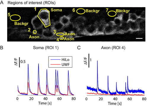
HiLo microscopy enhances calcium signals. (A) In vivo HiLo image of motor neurons of a zebrafish embryo expressing GCaMP5G in soma (ROI1), axons (ROIs 2–4), or background (ROIs 5–7). Scale bar 5 μm. (B) ΔF/F time series in the soma (ROI 1) shows that HiLo mode leads to a larger signal than UWF mode. (C) For axons (ROI 4) the gain in HiLo mode is even larger. Acquisition rate was 25fps. |
Expression Data
Expression Detail
Antibody Labeling
Phenotype Data
Phenotype Detail
Acknowledgments
This image is the copyrighted work of the attributed author or publisher, and
ZFIN has permission only to display this image to its users.
Additional permissions should be obtained from the applicable author or publisher of the image.
Full text @ PLoS One