
Fig. 2
- ID
- ZDB-FIG-171101-5
- Publication
- Lauterbach et al., 2015 - Fast Calcium Imaging with Optical Sectioning via HiLo Microscopy
- Other Figures
- All Figure Page
- Back to All Figure Page
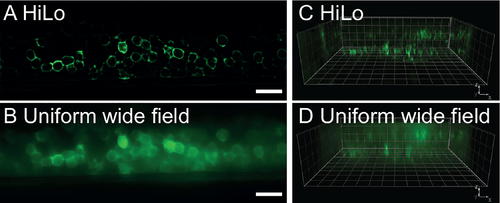
Optical section in embryonic spinal cord of zebrafish obtained with HiLo microscopy. (A) Lateral view of the spinal cord of transgenic zebrafish embryo expressing the fluorescent protein GCaMP5 pan-neuronally in Tg(elavl3:GCaMP5G) obtained with HiLo microscopy. (B) Same image obtained in UWF mode. The spatial distribution of GCaMP5G remains hidden under a haze of background fluorescence due to missing optical sectioning.Scale bars in both panels are 20 μm. (C) Corresponding z-stack in 3D visualization, imaged in HiLo mode. The pools of neurons on each side of the spinal cord are visible in two separate planes. (D) Same reconstruction in WF mode (grid step 10 μm/line). See also S1 Movie. |