
Fig. S6
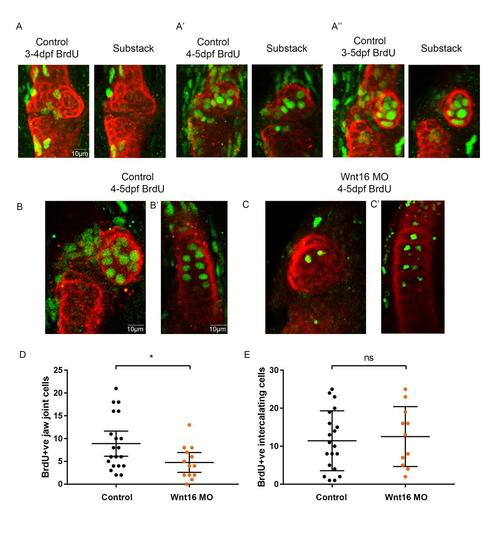
BrdU staining reveals cells proliferation at the jaw joint is affected by Wnt16 morpholino knockdown. (A-A''): Cell proliferation at the jaw joint occurs frequently between 4-5dpf. Pulse-chase experiments exposing control zebrafish to BrdU between 3-4dpf (A), 4-5dpf (A’) and 3-5dpf (A''). (A-A'') include max projection of image and substack through the cartilage joint. Anti-BrdU (green) and anti- collagen-II (red) label proliferating cells and cartilage at the jaw joints, respectively. (B-C): Pulse- chase experiments exposing control (B,B’) and Wnt16 MO injected (C,C') zebrafish to BrdU between 4-5dpf. Anti-BrdU (green) and anti-collagen-II (red) label proliferating cells and cartilage, respectively, at the jaw joint (left panel) and intercalating MC element region (right panel). (D): Number of BrdU positively labelled cells (BrdU +ve) at the jaw joint of 5dpf control injected and Wnt16 MO injected zebrafish after exposure to BrdU between 4-5dpf. (n=20, 13 joints). (E): Number of BrdU positively labelled cells (BrdU +ve) in the mid MC element intercalating region of 5dpf control injected and Wnt16 MO injected zebrafish after exposure to BrdU between 4-5dpf. (n=20, 11 joints). A Mann-Whitney U test was performed for (D) and a two-tailed student t-test for (E). ns= not significant, *=p≤0.05, **=p≤0.01, ***=p≤0.001. Bars on graph represent mean and 95%CI. |