
FIGURE
Fig. S7
- ID
- ZDB-FIG-170907-52
- Publication
- Gao et al., 2017 - The signalling receptor MCAM coordinates apical-basal polarity and planar cell polarity during morphogenesis
- Other Figures
- All Figure Page
- Back to All Figure Page
Fig. S7
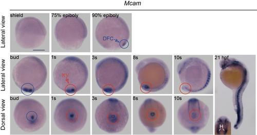
The spatiotemporal distribution ofmcam mRNAin zebrafish embryos. Zebrafish mcam mRNA expression was measured in DFCs (blue circles) and in KV (red circles) of wild-type embryos. At 21 hour post fertilization (hpf), mcam was expressed at blood vasculature, heart, somite, and eye field. The inset indicates expression of mcam mRNA in the heart. Inset: H, heart. Scale bar, 250 μm. |
Expression Data
| Gene: | |
|---|---|
| Fish: | |
| Anatomical Terms: | |
| Stage Range: | Shield to 20-25 somites |
Expression Detail
Antibody Labeling
Phenotype Data
Phenotype Detail
Acknowledgments
This image is the copyrighted work of the attributed author or publisher, and
ZFIN has permission only to display this image to its users.
Additional permissions should be obtained from the applicable author or publisher of the image.
Full text @ Nat. Commun.