
Fig. 6
- ID
- ZDB-FIG-170907-50
- Publication
- Gao et al., 2017 - The signalling receptor MCAM coordinates apical-basal polarity and planar cell polarity during morphogenesis
- Other Figures
- All Figure Page
- Back to All Figure Page
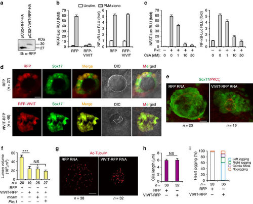
MCAM controls lumenogenesis by activation of NFAT. (a) Detecting the expression of RFP-HA and VIVIT-RFP-HA in HEK293 cells by IB. (b) VIVIT selectively inhibiting NFAT reporter activity. Jurkat cells were co-transfected with NFAT-Luc (left panel) or NF-kB-Luc (right panel) reporter plasmid, and with RFP and RFP-VIVIT expression plasmids. Twenty-four hours after transfection, cells were left untreated or were stimulated for 6 h with phorbol 12-myristate 13-acetate (PMA; 20 nM) and ionomycin (1 mM) (P+I). (c) Calcineurin dependence of NFAT and NF-kB reporter activity. Jurkat cells were transfected with NFAT-Luc (left panel) or NF-kB-Luc (right panel) reporter plasmid. Twenty-four hours after transfection, cells were left unstimulated or were stimulated for 6 h with P+I in the absence or presence of 1 μM cyclosporin (CsA). (d) Both RFP-tag and VIVIT-RFP were localized and expressed in DFC after microinjection of RFP and RFP-VIVIT mRNA into DFC of zebrafish KV. DIC means digital image of contrast. (e,f) Messenger RNAs were injected into zebrafish DFC of Sox17:GFP transgenic embryos, which were harvested at the 10 s stage. Lumen cells were labelled with an antibody against aPKCζ (red). Data are presented as mean±s.e.m. One-way ANOVA with Tukey’s post-test. ***P value<0.001 and NS=not significant. (g,h) KV cilia were labelled with an antibody against acetylated tubulin after injection of the indicated mRNAs. Data are presented as mean±s.e.m. and analysed using unpaired student’s t-test. The NS means not significant. (i) Quantitative analysis of heart joggings after injection of the indicated mRNAs into DFC of Sox17:GFP zebrafish embryos. Normal (left), reversed (right) and absent (no) jogging were calculated. Scale bar, 20 μm. n, number of observed embryos (e-i). |