
FIGURE
Fig. S8
- ID
- ZDB-FIG-170907-43
- Publication
- Gerri et al., 2017 - Hif-1α regulates macrophage-endothelial interactions during blood vessel development in zebrafish
- Other Figures
- All Figure Page
- Back to All Figure Page
Fig. S8
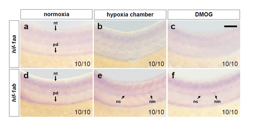
hif-1aa and hif-1ab are not highly expressed in ECs at 54 hpf. (a-c) Brightfield images of WISH for hif-1aa expression in 54 hpf WT embryos in normoxia, after hypoxia chamber or DMOG treatment for 6 hours starting at 48 hpf; lateral views. (d-f) Brightfield images of WISH for hif-1ab in 54 hpf WT embryos in normoxia, after hypoxia chamber or DMOG treatment for 6 hours starting at 48 hpf; lateral views. nt: neural tube; pd: pronephric duct; nc: notochord; nm: neuromast. n = 10 embryos from 3 different clutches. Scale bar, 50 μm. |
Expression Data
Expression Detail
Antibody Labeling
Phenotype Data
Phenotype Detail
Acknowledgments
This image is the copyrighted work of the attributed author or publisher, and
ZFIN has permission only to display this image to its users.
Additional permissions should be obtained from the applicable author or publisher of the image.
Full text @ Nat. Commun.