|
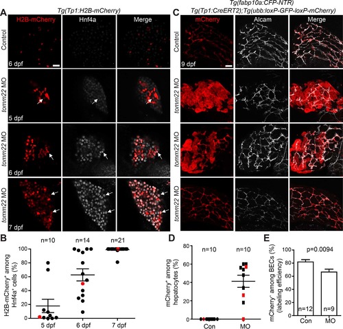
Biliary epithelial cells (BECs) give rise to hepatocytes in tomm22 MO-injected larvae. (A) Confocal single-optical section images showing Tp1:H2B-mCherry (red) and Hnf4a (gray) expression in the liver. Arrows point to H2B-mCherry/Hnf4a double-positive cells. (B) A graph showing the percentage of H2B-mCherry+ cells among Hnf4a+ cells in the livers of the tomm22 MO-injected larvae. Red dots indicate the larvae shown in (A). (C) Confocal projection images showing the hepatic expression of ubb:mCherry (red, Cre-labeled cells) and Alcam (gray, BECs) at 9 dpf. 4-OHT was treated from 48 to 84 hpf. (D) Graph showing the percentage of ubb:mCherry+ hepatocytes, which were derived from BECs. fabp10a:CFP-NTR expression was used to define hepatocytes. Red marks indicate the larvae shown in (C). (E) Graph showing the percentage of mCherry+ cells among Alcam+ BECs at 9 dpf, indicating Cre-mediated labeling efficiency. n indicates the number of larvae examined. Scale bars: 20 μm; error bars: ±SEM.
|