
Fig. S4
- ID
- ZDB-FIG-170706-26
- Publication
- Pei et al., 2016 - Additive reductions in zebrafish PRPS1 activity result in a spectrum of deficiencies modeling several human PRPS1-associated diseases
- Other Figures
- All Figure Page
- Back to All Figure Page
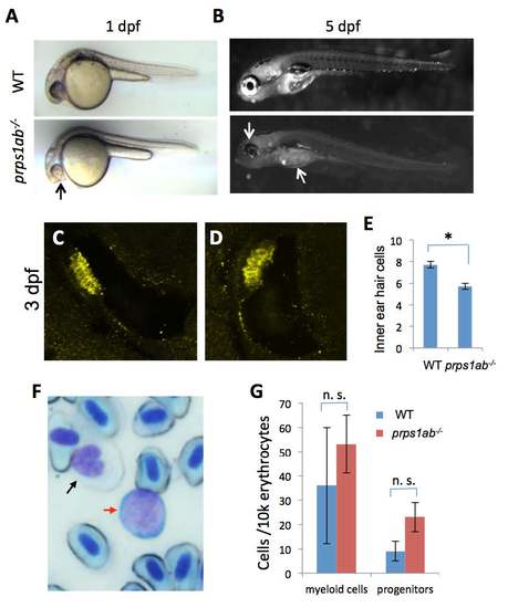
prps1a;prps1b double mutant phenotypes. (A) Reduced melanocyte pigmentation in the double mutant at 1 dpf. prps1ab-/- indicates prps1a;prps1b double mutant. Black arrow points to the small and poorly-pigmented eye of the double mutant. (B) Reduced iridophores in the double mutants at 5 dpf. White arrows indicate the reduced iridophores in the eye and yolk of the double mutant. (C-D) Inner ear hair cells in 3 dpf embryos were stained with a combination of myosin-VIIa and hair cell soma-1 antibodies. 11 embryos per group were used for inner ear hair cell analysis. Representative images are shown. (E) Quantification of the reduction of inner ear hair cells. The reduction in the double mutants is significant (n=11, p<0.003). (F) Blood cell lineage analysis by Wright Giemsa staining. Black arrow indicates a myeloid cell and red arrow indicates a progenitor cell. Other cells are erythrocytes. (G) Quantification of myeloid cells and progenitors in the 3 control adults and 3 double mutant adults at 4 months. The differences were not significant (n. s.). |
| Fish: | |
|---|---|
| Observed In: | |
| Stage Range: | Prim-5 to Adult |