
Fig. S2
- ID
- ZDB-FIG-170706-24
- Publication
- Pei et al., 2016 - Additive reductions in zebrafish PRPS1 activity result in a spectrum of deficiencies modeling several human PRPS1-associated diseases
- Other Figures
- All Figure Page
- Back to All Figure Page
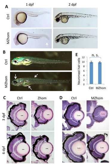
prps1a mutant phenotypes. (A) The smaller eye phenotype in prps1a MZhom embryos at 1 dpf and 2 dpf. White arrow indicates easily visible reduction in size. (B) Reduced iridophores in prps1a MZhom at 5 dpf. White arrows show where the reduction of iridophores in the head, trunk and yolk are most easily seen. (C) Retinal morphology of prps1a Zhom embryos at 3 dpf and 6 dpf. (D) Retinal morphology of prps1a MZhom embryos at 3 dpf and 6 dpf. CMZ, ciliary marginal zone. GCL, ganglion cell layer. IPL, inner plexiform layer. INL, inner nuclear layer. OPL, outer plexiform layer. ONL, outer nuclear layer. PR, photoreceptor. RPE, retina pigment epithelium. Eyes are most reduced at 3 dpf, but begin to recover by day 6. (E) prps1a homozygous mutants have a normal number of neuromast hair cells at 5 dpf. The difference between the control and mutants is not significant (n=10, p=0.87). |
| Fish: | |
|---|---|
| Observed In: | |
| Stage Range: | Prim-5 to Day 6 |