
FIGURE
Fig. S3
- ID
- ZDB-FIG-170706-25
- Publication
- Pei et al., 2016 - Additive reductions in zebrafish PRPS1 activity result in a spectrum of deficiencies modeling several human PRPS1-associated diseases
- Other Figures
- All Figure Page
- Back to All Figure Page
Fig. S3
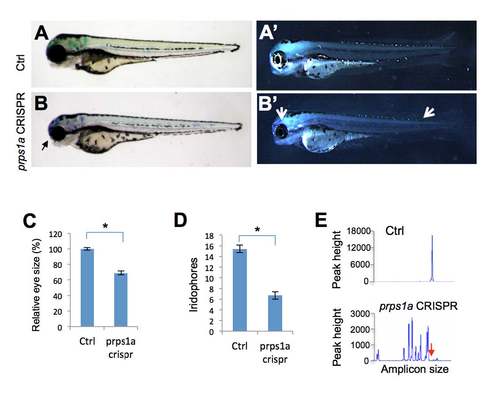
prps1a CRISPR mutant phenotypes. (A-B) Small eye phenotype in prps1a CRISPR mutants at 3 dpf. Black arrow points to the small eye in the mutant. (A’-B’) Iridophore phenotype in prps1a CRISPR mutants at 3 dpf. Two white arrows point to the reduced iridophores in the eye and dorsal trunk of the mutant. (C-D) Quantification of the reduction in eye size (C) and iridophores (D). The eye areas were calculated using image J. The numbers of iridophores were obtained by counting the iridophores in the dorsal and ventral side of the trunk area above the yolk extension. The reduction was significant in the eye size (n=10, p<0.001) and iridophores (n=10, p<0.001). (E) Representative fluorescent PCR plots showing the peaks indicating efficient somatic mutation. The representative plots are from Control # 4 and prps1a CRISPR #6 embryos (details in Suppl. Table 1). The X-axis represents the size of the target amplicon. The Y-axis shows the peak height for each amplicon size. The red arrow points to the position of the WT peak. All peaks shown are within 50 base pair of the WT peak. Ctrl is uninjected sibling fish. |
Expression Data
Expression Detail
Antibody Labeling
Phenotype Data
| Fish: | |
|---|---|
| Knockdown Reagent: | |
| Observed In: | |
| Stage: | Protruding-mouth |
Phenotype Detail
Acknowledgments
This image is the copyrighted work of the attributed author or publisher, and
ZFIN has permission only to display this image to its users.
Additional permissions should be obtained from the applicable author or publisher of the image.
Full text @ Sci. Rep.